Ureteral stricture is a section of the ureter channel that carries the urine produced in the kidney to the bladder becoming narrower than normal and the urine flow being obstructed. While the passage of urine can partially continue when the stricture is mild, an advanced-degree obstruction can cause urine to accumulate in the kidney, the kidney to swell and the kidney functions to be damaged over time.
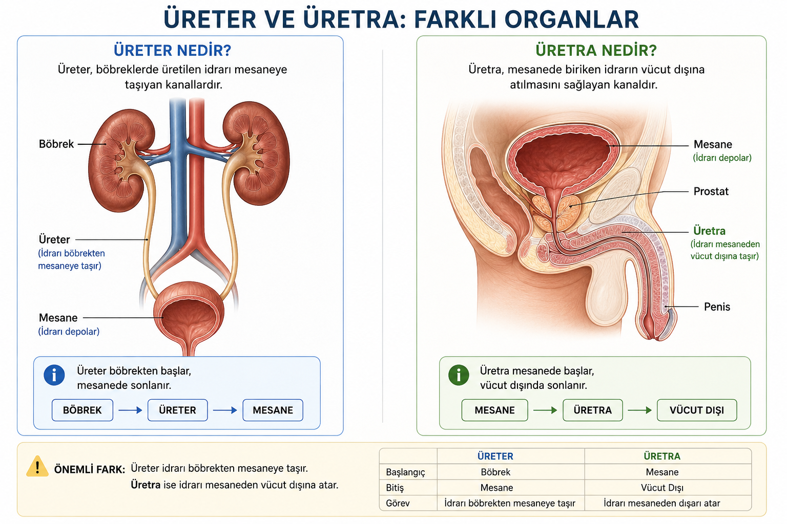
Another condition frequently confused with ureteral stricture is urethral stricture. Although their names resemble one another, the ureter and the urethra are different anatomical structures. Therefore ureteral stricture and urethral stricture are not the same disease. While ureteral stricture affects the urine flow between the kidney and the bladder, urethral stricture obstructs the urine flow that takes place from the bladder to outside the body. Their symptoms, causes and the treatments that can be applied are also different from one another.
The development of a complete or advanced-degree obstruction in the ureter in particular is important for the kidney. While severe flank pain arises in some patients, in some patients the stricture can progress for a long time without creating a marked complaint. For this reason, in treatment the aim is not only the relief of the patient's pain but also ensuring that the urine reaches the bladder from the kidney comfortably and the preservation of the kidney function.
What Is the Ureter?
The ureter is the thin, tube-shaped organ with a muscular structure that provides the transport of the urine formed in the kidney to the bladder. Normally there are two ureters, one belonging to the right and the other to the left kidney. The ureters start from the renal pelvis region where the kidney collects the urine and extend as far as the bladder.
The urine does not advance to the bladder only thanks to gravity. The rhythmic contractions created by the smooth muscles in the ureter wall, that is, peristaltic movements, help the urine to be carried from the kidney towards the bladder.
When a narrowing or obstruction occurs at any point of the ureter, this system can be disrupted. When the passage of the urine to the bladder becomes difficult, the pressure in the upper part of the stricture begins to increase. This pressure can be reflected in the urine collecting system of the kidney and cause the dilatation named hydronephrosis.
What Is the Urethra?
The urethra is the channel that provides for the urine accumulated in the bladder to be expelled outside the body. The anatomy of the urethra is different in women and men. The urethra, which is shorter in women, opens to the outside of the body in men by passing through the prostate and the penis.
Therefore, by simplifying the path the urine follows in the urinary system we can show it as follows:
- Kidney: Produces the urine.
- Ureter: Carries the urine from the kidney to the bladder.
- Bladder: Stores the urine.
- Urethra: Provides for the urine in the bladder to be expelled from the body.
This anatomical distinction makes it easier to understand why ureteral stricture and urethral stricture are not the same disease.
Are Ureteral Stricture and Urethral Stricture the Same?
No. Ureteral stricture and urethral stricture are different diseases. In ureteral stricture the problem is in the channel between the kidney and the bladder. In urethral stricture, however, there is a narrowing in the urinary channel between the bladder and the outside of the body.
For this reason the symptoms patients experience can also differ. In ureteral stricture, flank or side pain due to the increase in pressure in the kidney, hydronephrosis, recurrent infections and deterioration in kidney function can be to the fore. In urethral stricture, however, lower urinary tract symptoms such as weakening of the urine flow, urinating intermittently, urinating by straining, not being able to empty the bladder completely and the prolongation of the urination time are seen more frequently.
Why Does Ureteral Stricture Occur?
Ureteral strictures can be congenital as well as develop later. In strictures that appear later, injury to the ureter wall, inflammation or the formation of scar tissue during healing can reduce the diameter of the channel.
The main conditions that can lead to ureteral stricture are the following:
- Ureteral or kidney stone disease
- The formation of long-term obstruction because of a ureteral stone
- Endoscopic interventions carried out for a ureteral stone
- Previously undergone urinary system operations
- Some surgical procedures carried out in the abdomen and pelvis region
- Injuries occurring in the ureter
- Tissue changes that can develop after radiotherapy
- Infection and chronic inflammation
- Diseases affecting the ureter from outside such as retroperitoneal fibrosis
- Masses pressing on the ureter from outside
- Tumours of the ureter and its surroundings
- Congenitally developed urinary system strictures
The ureteral stone remaining in the same region for a long time in particular can cause inflammation and damage in the tissue around the stone. Similarly, the tissue healing that develops after some endoscopic procedures can also rarely create a stricture.
What Are the Symptoms of Ureteral Stricture?
The symptoms of ureteral stricture can vary according to the degree and location of the obstruction, how quickly it has developed and whether it is one-sided or two-sided. Slowly developing strictures may not give symptoms for a long time in some patients.
Among the symptoms that may be seen, the following may be found:
- Pain in the lower back or side region
- Intermittent or continuous pain on the flank
- Recurrent urinary tract infections
- Fever and shivering
- Nausea and vomiting
- Blood being seen in the urine
- Dilatation in the kidney, that is, hydronephrosis
- Deterioration in kidney functions
In some patients in whom the stricture develops slowly there may be no pain. The problem may be noticed by dilatation being seen in the kidney during an ultrasonography or computed tomography performed for another reason. For this reason the absence of pain does not mean that the obstruction in the ureter is unimportant.
Does Ureteral Stricture Damage the Kidney?
Yes, a ureteral stricture that continues for a long time in particular and that markedly obstructs the urine flow can affect the kidney function. When the ureter is obstructed, the urine that continues to be produced in the kidney cannot empty sufficiently into the bladder. The pressure in the upper part of the stricture rises and the collecting system of the kidney begins to dilate.
This situation continuing for a long time can reduce the function of the kidney tissue. If infection is also added to the picture, the situation can become more serious. For this reason one of the basic aims in ureteral stricture is the preservation as far as possible of the existing kidney function.
How Is Ureteral Stricture Diagnosed?
In the diagnostic process not only whether there is a narrowing in the ureter but also the location, length and degree of the stricture and its effect on the kidney are evaluated. This is one of the basic factors determining the choice of treatment.
According to the patient's condition, the following examinations can be made use of:
- Ultrasonography
- Computed tomography and, when necessary, CT urography
- Magnetic resonance imaging and MR urography
- Blood tests and evaluation of the kidney functions
- Urine test and, when necessary, urine culture
- Retrograde pyelography
- Antegrade imaging methods
- Ureteroscopic evaluation
- In necessary patients, nuclear medicine examinations that evaluate the function and drainage of the kidneys separately
Knowing how much the kidney is working in particular is important in treatment planning. The treatment approach of a patient who has an advanced-degree stricture but whose kidney function is preserved and of a patient with a kidney that has been obstructed for a long time and whose function has decreased seriously may not be the same.
How Is Ureteral Stricture Treated?
Ureteral stricture treatment is planned specifically for the patient. The location and length of the stricture, the treatments previously applied, the cause of the stricture, the function of the kidney and the patient's general health status directly affect the choice of treatment.
The basic aims of the treatment are the following:
- To re-establish the urine flow from the kidney to the bladder
- To reduce the high pressure formed in the kidney
- To preserve the kidney function
- To prevent recurrent infections
- To ensure that the stricture is eliminated as permanently as possible
Placing a Ureteral Stent
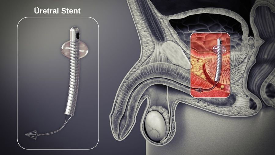
The ureteral stent is a thin tube placed between the kidney and the bladder. One end of the frequently used double-J or DJ stents remains in the kidney, the other end in the bladder. In this way a channel is created through which the urine can pass through the narrowed region.
In some patients the stent is used for the purpose of temporarily providing the drainage of the kidney. In infection, acute obstruction or before reconstructive surgery to be planned, it may be necessary to relieve the kidney.
However the stent is not always a permanent treatment that eliminates the stricture. Depending on the characteristics of the stricture, obstruction can develop again when the stent is removed. In patients who need to use a stent for a long time, the stent may need to be changed at certain intervals.
Percutaneous Nephrostomy
If the kidney needs to be emptied urgently because of the obstruction in the ureter, percutaneous nephrostomy can be applied. In this method, accompanied by imaging, a thin catheter is placed through the skin into the collecting system of the kidney and the urine is enabled to empty into a bag outside.
Especially in some situations in which the obstruction is accompanied by infection, rapid drainage of the kidney is important. Nephrostomy can at the same time be used as a temporary solution so that the subsequent treatment can be planned.
Endoscopic Ureteral Stricture Treatment
Some short ureteral strictures with appropriate characteristics can be treated with endoscopic methods. The stricture region is reached with thin devices advanced through the urinary tract and an attempt is made to widen the narrow segment.
Among the endoscopic treatments there may be methods such as balloon dilatation or endoureterotomy, which means the endoscopic cutting of the stricture tissue. After the procedure, a temporary stent can be used during the healing period in order to ensure that the ureter remains open.
The important advantage of endoscopic methods is that they do not require a large surgical incision. Nevertheless not every stricture is suitable for endoscopic treatment. In strictures that are long, of dense fibrotic structure, that have developed after radiotherapy or that have recurred despite having been treated before, reconstructive surgery may be more suitable.
Reconstructive Surgery in Ureteral Stricture
In patients in whom the permanent elimination of the stricture is aimed at, ureteral reconstruction can come onto the agenda. The aim of reconstructive surgery is not only to widen the narrow region but to create a path between healthy tissues through which the urine can pass comfortably.
Which operation will be applied depends on which part of the ureter the stricture is in and how long it is.
Removing the Narrow Part of the Ureter and Rejoining It
If there is a stricture in a short ureteral segment, the narrow and unhealthy part can be removed. Afterwards the healthy ends of the ureter can be rejoined in a way that does not create tension. This procedure is called ureteroureterostomy.
Reconnecting the Ureter to the Bladder
If the stricture is in the lower ureter section close to the bladder, after the problematic segment is removed the healthy ureter can be reconnected to the bladder. This operation is named ureteroneocystostomy or ureteral reimplantation.
In longer strictures in which the healthy end of the ureter cannot directly reach the bladder, a part of the bladder can be extended towards the ureter. This reconstruction is known as the Boari flap. In some patients the psoas hitch technique can also be made use of to bring the bladder closer to the ureter.
The Treatment of Longer and Complex Ureteral Strictures
Ureteral strictures affecting a long segment or recurring despite having been treated before may require more complex reconstruction. According to the patient's anatomy and the characteristics of the stricture, the use as a graft of mucosal tissue taken from inside the mouth, the reconstruction of the ureter with a segment from the intestine or different reconstructive methods can be evaluated.
In this type of operation the aim is to preserve the kidney as far as possible and to create a low-pressure, continuous urinary path between the kidney and the bladder.
Ureteral Stricture Treatment with Robotic Surgery
In suitable patients ureteral reconstruction can be carried out with robotic surgery. Robotic systems providing a magnified three-dimensional image and the surgical instruments allowing precise movements in narrow areas offer the surgeon important technical possibilities especially in reconstructive operations that require the rejoining of thin structures such as the ureter.
With the robotic approach, ureteroureterostomy, the reconnection of the ureter to the bladder, psoas hitch, the Boari flap and selected complex reconstructions can be applied. However the choice of the operation method should be made not only according to the technology being available but according to the anatomy of the stricture and the characteristics of the patient.
In robotic or laparoscopic minimally invasive approaches, being able to perform the operation with smaller incisions compared with open surgery can provide an advantage in suitable patients in terms of post-operative pain and the recovery process.
What Happens If Ureteral Stricture Is Not Treated?
Leaving a stricture that significantly obstructs the passage of urine untreated can, depending on the degree and duration of the stricture, lead to increasing dilatation and loss of function in the kidney. Problems such as recurrent urinary tract infections and stone formation can also be seen.
Infection accompanying an obstructed kidney in particular may require urgent evaluation. In patients with fever, shivering, severe flank pain and deterioration in general condition, medical help should be obtained without delay.
Why Does Urethral Stricture Occur?
Urethral stricture is the development of scar tissue in the urethral channel that carries urine out from the bladder and the narrowing of the channel. Because the urethra is longer in men, it is an important urological problem encountered more frequently clinically.
Among the causes of urethral stricture there may be previously undergone urethral interventions, catheterisation, trauma, infection, pelvic injuries and some surgical procedures. In some patients, however, a definite cause may not be able to be determined.
What Are the Symptoms of Urethral Stricture?
- Weakening of the urine flow
- The need to strain while urinating
- The urine coming thin or scattered
- Voiding taking a long time
- The feeling that the bladder is not emptying completely
- Frequent urination
- Recurrent urinary tract infections
- In advanced situations, being unable to urinate
As can be seen, the symptoms of urethral stricture arise mostly during the expulsion of the urine out from the bladder. In ureteral stricture, however, the basic problem is that the urine formed in the kidney cannot reach the bladder.
Which Method Is Preferred in Ureteral Stricture Treatment?
One of the questions patients ask most frequently is which of a stent, an endoscopic intervention or an operation is better in ureteral stricture. It is not correct to define a single method as the best treatment for all patients.
In some strictures that are short and of suitable structure, endoscopic treatment can be considered. In a situation of acute obstruction or infection, the drainage of the kidney with a stent or nephrostomy may be needed first. In long, recurrent or complex strictures, however, reconstructive surgery can be evaluated for the purpose of a more permanent solution.
For this reason it is important that answers be sought to four questions before a treatment decision is made: Where is the stricture? How long is it? How much is the kidney working? What is the cause of the stricture? After this information is obtained, the suitable one of the endoscopic, open, laparoscopic or robotic reconstructive methods can be chosen.
Frequently Asked Questions About Ureteral Stricture
Does ureteral stricture go away on its own?
A permanent ureteral stricture due to genuine scar tissue is not expected to correct itself completely. However the cause and degree of the obstruction must definitely be evaluated with imaging and the necessary function tests.
Is ureteral stricture dangerous?
An advanced-degree ureteral stricture or one that continues for a long time is important because it can cause an increase in pressure in the kidney and loss of function. Infection accompanying the obstruction in particular may require urgent treatment.
Does ureteral stricture cause pain?
Yes. Pain can be seen in the lower back and flank region. However, since some chronic strictures can progress without creating marked pain, an evaluation should not be made only according to the presence of pain.
How long does the stent stay in ureteral stricture?
The duration of use of the stent varies according to the reason for which it is applied and the planned treatment. The duration of stay of a stent used for the purpose of temporary drainage and of a stent used after reconstructive surgery may not be the same. The duration must be determined by the urologist following up the patient.
Is ureteral stricture surgery difficult?
The degree of difficulty of the operation varies according to the location and length of the stricture, the tissues around it, previously undergone surgeries and the reconstruction method to be applied. The treatment of a short stricture and the reconstruction of a long ureter that has been operated on before are not of the same scope.
Does ureteral stricture recur after the operation?
The possibility of the stricture recurring can vary according to the method used, the length and cause of the stricture and the characteristics of the tissue. For this reason it is important that imaging and the kidney functions be followed up at certain intervals after the treatment.
What is the simplest difference between ureteral stricture and urethral stricture?
The ureter carries urine from the kidney to the bladder, and the urethra from the bladder to outside the body. For this reason ureteral stricture and urethral stricture are two different urological diseases affecting, respectively, the emptying of the kidney and the emptying of the bladder to the outside.
Can the kidney be saved in ureteral stricture?
The answer to this question depends on the existing function of the kidney and the duration and severity of the obstruction. If there is sufficient function in the kidney, the aim is the preservation of the kidney with treatments that will re-establish the urine flow. In advanced loss of function, however, the treatment decision is made by evaluating the remaining function of the kidney and the patient's general condition together.
Clinical Note
This article was written and medically reviewed by Prof. Dr. Murat Binbay, a urologist with more than 25 years of experience and over 1,500 robotic operations. The assessments reflect current international guideline recommendations together with our own clinical practice.
The information here is for general guidance only. Because every patient's history, imaging findings and comorbidities differ, a treatment decision can only be made after an examination. You can reach us through our contact page, or read more about our physician on the about us page.
Sources
- European Association of Urology — Urethral Strictures Guidelines
- European Association of Urology — Clinical Guidelines
Last reviewed: 8 September 2026 · Reviewed by: Prof. Dr. Murat Binbay, Urology and Robotic Surgery Specialist
Let Prof. Dr. Murat Binbay's team review your case
If you read this far, you probably have questions. Fill in the two-minute digital consultation form and we will get back to you within 24 hours.
- Answer within 24 hours
- Your data stays confidential

Urology and Robotic Surgery Specialist · 25+ years of experience
Last medically reviewed:
 Önceki Yazı
What Is Urinary Incontinence? Its Causes, Types and Treatment
Önceki Yazı
What Is Urinary Incontinence? Its Causes, Types and Treatment
 Sonraki Yazı
Neurogenic Bladder
Sonraki Yazı
Neurogenic Bladder
Yorumlar
Yorum Yazın
Henüz yorum yapılmamış. İlk yorumu siz yapın!