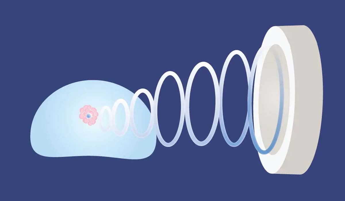
Prostate biopsy is the procedure of taking small pieces of prostate from the prostate gland. The pieces taken with the biopsy are stained with special dyes and examined under the microscope. The pathologist examining these pieces determines whether the patient has prostate cancer; and if there is prostate cancer, how aggressive the cancer is is also determined thanks to the prostate biopsy.
A definitive prostate cancer diagnosis is made thanks to prostate biopsy.
Types of Prostate Biopsy
Prostate biopsy is performed in 2 different types.
The Old Type: Classic Prostate Biopsy
Classic-type prostate biopsy can be applied in the urology department of almost every hospital.
In the classic type of biopsy only transrectal ultrasonography is used. That is, there is no MRI examination procedure before the biopsy.
With the ultrasound probe extended through the patient's rectum, 12 pieces are taken from the prostate at random. During this procedure the urology specialist carries out the procedure without seeing the cancerous areas (if any) in the prostate.
The New Type: Prostate Fusion Biopsy
With prostate fusion biopsy, currently applied in a limited number of hospitals in our country, aggressive prostate cancer can be detected exactly.
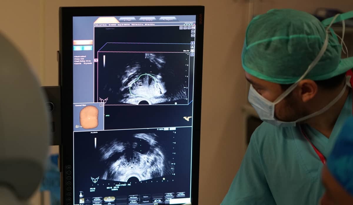
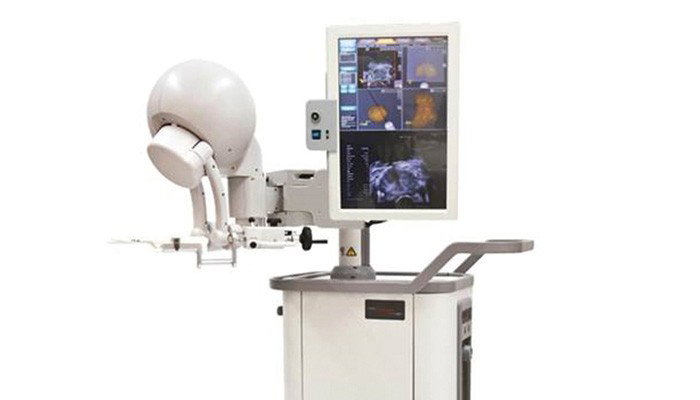
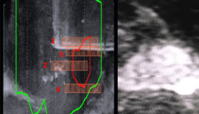
Before the procedure a multiparametric prostate MRI is taken of the patient. The areas appearing suspicious for cancer are marked on the MRI. Afterwards, entry is made with an ultrasound probe through the anus of the patient, who has been put to sleep with anaesthesia. With the aid of the biopsy machine the MRI images and the ultrasound images are matched. Cancerous regions that neither one could see are clearly identified with this method.
The procedure of taking a sample from the prostate is not done at random. Samples are taken from regions carrying a risk of cancer and a high rate of cancer detection is achieved.
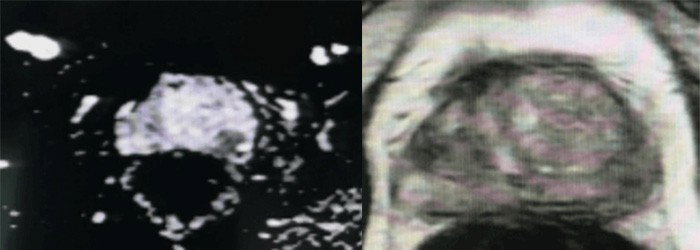
| Classic Prostate Biopsy | Prostate Fusion Biopsy |
| No MRI is taken of the patient before the prostate biopsy. | A multiparametric prostate MRI is taken of the patient before the prostate biopsy. |
| Generally the patient is not put to sleep and local anaesthesia is applied to the patient. | Generally the procedure is carried out under anaesthesia with the patient put to sleep. |
| The patient generally feels pain. | The patient feels no pain. |
| Particularly rapidly spreading aggressive prostate cancers can be overlooked. | Particularly rapidly spreading aggressive prostate cancers are not overlooked. |
| Treatment planning may be done incorrectly. | Treatment planning is done correctly. |
In Which Situations Should a Prostate Biopsy Be Performed?
In order to make a definitive diagnosis of prostate cancer, performing a prostate biopsy on people at risk of cancer is the most logical approach.
We can list the people at risk in terms of prostate cancer as follows:
- Men with a high PSA level in the blood
- Those who have an asymmetric prostate on digital rectal examination or men with hardness or a mass on the surface of the prostate
- Men in whom a suspicious area for cancer has been detected within the prostate on MRI imaging
When a prostate biopsy is performed only from the risky regions imaged on MRI, as much as 10% of the cancerous regions cannot be detected. For this reason it is recommended that multiparametric MRI imaging also be used.
The Application of Prostate Fusion Biopsy
Preparation for the Biopsy
Before the prostate fusion biopsy an MRI is taken of the patient.
During the procedure the patient is put to sleep with anaesthesia. Eating and drinking need to be stopped 6-8 hours before the anaesthesia.
Before the biopsy, certain tests are carried out to make sure that the patient does not have an untreated urinary tract infection.
Because the biopsy procedure is performed through the anus, the bowels are cleared beforehand with an enema procedure.
Giving the patient antibiotics before the biopsy is also among the recommended procedures.
About 1 week before having a prostate fusion biopsy, the use of blood-thinning medicines needs to be limited. For this reason you must without fail state to your urology specialist the medicines you use.
The Biopsy Application
In the prostate biopsy procedure an ultrasound probe is placed into the patient's anus. The front, back and position of the prostate are determined.
The prostate pieces to be taken for examination can be taken by the method of perforating the large intestine. This route is called transrectal prostate biopsy.
Transrectal prostate biopsy: a transrectal ultrasound is placed into the large intestine through the anus of the patient, who is laid on their side. The needle extended with the transrectal ultrasound rapidly reaches the prostate and the procedure of taking sample pieces for examination is carried out. 10-12 pieces are taken from different points of the prostate. The procedure lasts on average 25 minutes.
Transperineal prostate biopsy: transperineal biopsy is not a very frequently used method. The patient is laid on their back or on their side. Before the procedure the patient's skin is sterilised.
A finger is inserted through the patient's rectum to hold the prostate. A needle is inserted from the perineal region and pushed into the prostate. The pieces to be taken for examination are taken by rotating the needle. The procedure lasts on average 30 minutes.
After the Biopsy
After the prostate biopsy the patient needs to rest for on average 6-8 hours.
The patient may have pain in the pelvic region.
Blood may be seen in the urine for 4-5 days following the procedure. In the same way, blood may also be seen in the semen for the following 6 weeks.
After the transrectal biopsy procedure there may be bleeding from the patient's anus.
When the procedure of taking antibiotics before the prostate fusion biopsy is not carried out, there is a risk of acute prostatitis (prostate infection) developing.
Can the Application Be Repeated?
Despite the first prostate biopsy result coming back as "benign prostatic hyperplasia or inflammation of the prostate", in the event of an increase in PSA values in the patient's follow-ups or of an abnormal finding on digital rectal examination, a repeat of the biopsy may be necessary.
In the event of the first prostate biopsy result coming back as "atypical small acinar proliferation (ASAP)", the biopsy can be repeated.
In the situation where "high-grade PIN" is detected in at least 3 samples in the first prostate biopsy result, the biopsy can also be repeated.
The PI-RADS Classification
The European Society of Urogenital Radiology published in 2012 a classification called the "Prostate Imaging Reporting and Data System" (PI-RADS). This classification was updated in 2014 and continues to be updated.
This classification aims for radiologists to assess the lesions within the prostate according to certain criteria. In this way an attempt is made to bring standardisation to the assessments.
The multiparametric MRI images taken are examined by radiologists experienced in this subject. The lesions within the prostate are scored between 1 and 5.
For PI-RADS 1-2 lesions, it is recommended that a classic prostate biopsy be performed.
For PI-RADS 3 the patient's other findings are also examined and a decision on prostate biopsy is made accordingly.
For PI-RADS 4-5 the interpretation is made that the lesions are at high risk in terms of prostate cancer.
There are 3 different methods in MRI fusion biopsy.
Biopsy under MRI: a biopsy can be taken with high accuracy from the point identified on MRI imaging.
In this method a biopsy is not taken only from the points imaged on MRI. A systematic biopsy must also be performed. This procedure is also not much preferred because it takes a long time and is expensive.
Cognitive MRI biopsy: biopsy samples are taken from the points marked on the MRI.
In this method, small suspicious points can be overlooked.
Software-assisted MRI ultrasound fusion biopsy: the prostate is examined with ultrasound. The images taken from the MRI are combined with computer software. As a result a three-dimensional MRI image is created.
In this method the procedure of taking a sample from all suspicious lesions is carried out.
Clinical Note
This article was written and medically reviewed by Prof. Dr. Murat Binbay, a urologist with more than 25 years of experience and over 1,500 robotic operations. The assessments reflect current international guideline recommendations together with our own clinical practice.
The information here is for general guidance only. Because every patient's history, imaging findings and comorbidities differ, a treatment decision can only be made after an examination. You can reach us through our contact page, or read more about our physician on the about us page.
Sources
- European Association of Urology — Prostate Cancer Guidelines
- National Cancer Institute (NIH) — Prostate Cancer
- Türk Üroloji Derneği — Güncel Kılavuzlar
Last reviewed: 8 September 2026 · Reviewed by: Prof. Dr. Murat Binbay, Urology and Robotic Surgery Specialist
Let Prof. Dr. Murat Binbay's team review your case
If you read this far, you probably have questions. Fill in the two-minute digital consultation form and we will get back to you within 24 hours.
- Answer within 24 hours
- Your data stays confidential

Urology and Robotic Surgery Specialist · 25+ years of experience
Last medically reviewed:
Yorumlar
10Yorum Yazın
Henüz yorum yapılmamış. İlk yorumu siz yapın!