When bladder cancer is diagnosed, the first question patients and their families usually ask is: “What stage is it?” It is the right question, because in bladder cancer the stage is the single most important piece of information for deciding which treatment is needed, whether the bladder can be preserved and how follow-up should be organised. In particular, whether or not the tumour has reached the muscle layer of the bladder wall separates two very different treatment pathways. In this article we explain in plain language the stages of bladder cancer, how the stage is determined and which treatment options are discussed at each stage.
The Structure of the Bladder Wall: the Key to Understanding Staging
The bladder is an organ that stores urine and has a layered wall. From the inside outwards there is a thin lining in contact with the urine (the urothelium), beneath it a layer of loose connective tissue (the lamina propria), then the thick muscle layer that contracts to empty the bladder (the detrusor muscle), and finally the fatty tissue surrounding the bladder.
The great majority of bladder cancers arise from the innermost urothelium. How deeply the tumour has grown through these layers is the main criterion that defines the stage of the disease. Roughly speaking, if the tumour has not reached the muscle layer it is called “non-muscle-invasive” (superficial) bladder cancer; if it has, it is called “muscle-invasive” bladder cancer. At diagnosis, about three in four patients fall into the non-muscle-invasive group.
Bladder Cancer Stages According to the TNM System
Bladder cancer is staged with the TNM system used worldwide. T describes the depth of the tumour in the bladder wall, N describes spread to the lymph nodes and M describes spread to distant organs (metastasis). The T categories in brief are:
- Ta: a tumour confined to the inner surface, growing into the bladder cavity (papillary), which has not invaded the underlying connective tissue.
- Tis (carcinoma in situ, CIS): a flat tumour that spreads along the surface but has aggressive cells. Although it looks limited, it has a high tendency to progress if left untreated.
- T1: the tumour has grown into the connective tissue beneath the lining (lamina propria) but has not reached the muscle layer.
- T2: the tumour has reached the muscle layer of the bladder (muscle-invasive disease).
- T3: the tumour has grown through the muscle into the fatty tissue around the bladder.
- T4: the tumour has spread into neighbouring organs such as the prostate, uterus or vagina, or to the pelvic or abdominal wall.
Ta, Tis and T1 tumours together form the “non-muscle-invasive bladder cancer” group. Tumours of T2 and above are considered “muscle-invasive bladder cancer”. If there is lymph node involvement (N1–N3) or distant metastasis (M1), the disease is regarded as advanced stage.
Stage and Grade Are Not the Same Thing

Alongside the stage, the pathology report also contains information on “grade”. The stage tells us how deeply the tumour has grown, while the grade describes how aggressive the cancer cells look under the microscope. In the current classification, tumours are divided into “low grade” and “high grade”. Low-grade tumours may recur often but are unlikely to progress into the muscle layer; high-grade tumours carry a greater risk of progression.
In non-muscle-invasive disease, stage and grade are assessed together to divide patients into low, intermediate, high and very high risk groups. The number and size of tumours, whether they have recurred before and whether CIS is present are also included in this risk assessment. The treatment plan is largely shaped by this risk group.
How Is the Stage Determined?
The first and most important step in staging bladder cancer is TUR-BT (transurethral resection of bladder tumour). In this procedure the tumour is removed through the urethra and sent for pathology. The operation has both a treatment and a diagnostic purpose. For accurate staging, the removed tissue must also contain the muscle layer beneath the tumour; otherwise it is not possible to tell whether the tumour has invaded the muscle. You can find detailed information in our article on TUR-T surgery in bladder cancer.
In some cases a second resection (re-TUR) is recommended a few weeks after the first. Particularly in T1 tumours, when the first specimen contains no muscle tissue or when the tumour could not be removed completely, a second procedure is important for detecting residual tumour that may have been missed and for confirming the stage.
In addition to the pathology assessment, imaging is also used. CT urography evaluates the kidneys and ureters as well, showing whether there are additional tumours in the upper urinary tract. In muscle-invasive disease, CT of the chest, abdomen and pelvis is used to assess the lymph nodes and distant organs. In selected patients, multiparametric bladder MRI can provide additional information on whether the tumour has invaded the muscle layer.
Treatment Approach in Non-Muscle-Invasive Bladder Cancer
In Ta, T1 and CIS tumours the main goals are to preserve the bladder, to reduce recurrence and to prevent progression into the muscle layer. Treatment starts with TUR-BT, after which medication instilled into the bladder is planned according to the risk group:
- In low-risk patients, a single dose of intravesical chemotherapy after surgery and regular check-ups are often sufficient.
- In intermediate-risk patients, a course of intravesical chemotherapy or BCG may be given.
- In high-risk patients, intravesical BCG with maintenance doses is the standard approach.
- In some very high-risk patients, early removal of the bladder (radical cystectomy) is also discussed with the patient as an option.
For more information on BCG and newly developed intravesical treatments, see our article on BCG and adenovirus-based treatments in superficial bladder cancers. Because tumours in this group tend to recur, regular follow-up cystoscopies are an integral part of treatment.
Treatment Approach in Muscle-Invasive Bladder Cancer
Once the tumour has reached the muscle layer (T2 and above), the behaviour of the disease changes. At this stage, simply resecting the tumour is not enough, because the risk of spread to the lymph nodes and distant organs rises markedly. In suitable patients, the standard approach is cisplatin-based chemotherapy before surgery (neoadjuvant chemotherapy) followed by removal of the bladder together with the lymph nodes (radical cystectomy). Randomised trials have shown that chemotherapy before surgery contributes to survival in suitable patients.
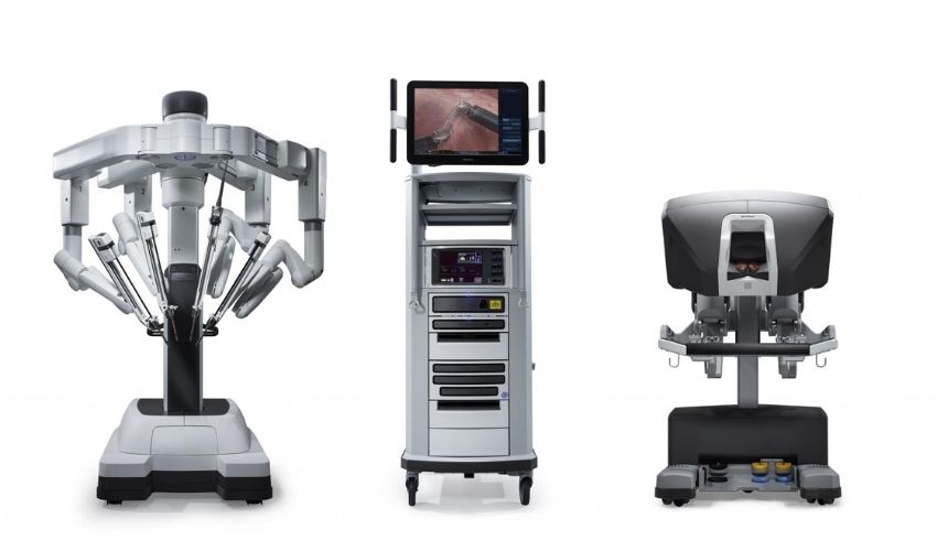
After the bladder is removed, a new route is created for urine to leave the body: either a new bladder made from bowel or a urinary opening on the abdominal wall (urostomy). Details on radical cystectomy and urinary diversion options can be found in our article on radical cystectomy surgery. In our clinic we perform these operations with robotic surgery; for the features of the robotic approach, see our article on robotic surgery for bladder cancer.
For selected patients who are not suitable for surgery or who wish to keep their bladder, “trimodal bladder-preserving therapy” — the most complete TUR-BT possible combined with chemotherapy and radiotherapy — is another option. Who is suitable for this approach is assessed by a multidisciplinary tumour board, taking into account the features of the tumour and the patient's general health.
Advanced and Metastatic Disease

When bladder cancer has spread to the lymph nodes or distant organs, systemic treatments form the basis of care. In recent years there has been significant progress in this field, with immunotherapy and targeted drugs alongside chemotherapy. Treatment of these patients requires medical oncologists, urologists and radiation oncologists to work together. At this stage surgery may be considered in selected patients, mostly to control symptoms.
Why Is Early Diagnosis So Important?
The most common symptom of bladder cancer is painless blood in the urine. Even if bleeding appears only once and then disappears, it must always be investigated, because catching the cancer at the non-muscle-invasive stage considerably increases the chance of preserving the bladder. We discuss this in detail in our article on blood in the urine. Smoking is the most important known risk factor for bladder cancer; stopping smoking both reduces the risk of new tumours and supports the treatment process.
Frequently Asked Questions
Is superficial bladder cancer a “mild” cancer?
Most non-muscle-invasive tumours can be well controlled; however, they have a strong tendency to recur and some may progress into the muscle layer over time. For this reason, treatment and regular check-ups should never be skipped.
Can the stage change during treatment?
The stage is determined by the situation at diagnosis. However, pathology after a second TUR-BT or radical cystectomy may show a more advanced stage than the first assessment. The treatment plan is therefore updated in the light of new information.
Does every patient need their bladder removed?
No. In the great majority of patients with non-muscle-invasive disease, the bladder is preserved. Removal of the bladder is mostly considered in muscle-invasive disease or in very high-risk superficial tumours.
Conclusion
In bladder cancer, the stage is the compass that sets the course of treatment. In tumours that have not reached the muscle layer the aim is to preserve the bladder while preventing recurrence and progression, whereas muscle-invasive disease requires more comprehensive treatment. Accurate staging depends on a high-quality TUR-BT, an experienced pathology assessment and appropriate imaging working together. We recommend discussing in detail with your doctor which stage your diagnosis is and what it means for you.
Clinical Note
This article was written and medically reviewed by Prof. Dr. Murat Binbay, a urologist with more than 25 years of experience and over 1,500 robotic operations. The assessments reflect current international guideline recommendations together with our own clinical practice.
The information here is for general guidance only. Because every patient's history, imaging findings and comorbidities differ, a treatment decision can only be made after an examination. You can reach us through our contact page, or read more about our physician on the about us page.
Sources
- Gontero P et al. EAU Guidelines on Non-muscle-invasive Bladder Cancer (TaT1 and CIS). European Association of Urology, 2024 edition.
- van der Heijden AG et al. EAU Guidelines on Muscle-invasive and Metastatic Bladder Cancer. European Association of Urology, 2024 edition.
- Brierley JD, Gospodarowicz MK, Wittekind C (eds). TNM Classification of Malignant Tumours, 8th edition. UICC, Wiley-Blackwell, 2017.
- Sylvester RJ et al. Predicting recurrence and progression in individual patients with stage Ta T1 bladder cancer using EORTC risk tables. Eur Urol 2006;49:466–477.
- Grossman HB et al. Neoadjuvant chemotherapy plus cystectomy compared with cystectomy alone for locally advanced bladder cancer. N Engl J Med 2003;349:859–866.
Last reviewed: 8 October 2026 · Reviewed by: Prof. Dr. Murat Binbay, Specialist in Urology and Robotic Surgery
Let Prof. Dr. Murat Binbay's team review your case
If you read this far, you probably have questions. Fill in the two-minute digital consultation form and we will get back to you within 24 hours.
- Answer within 24 hours
- Your data stays confidential

 Önceki Yazı
What Is a Double J Stent? Living With a Stent After Kidney Stone Treatment
Önceki Yazı
What Is a Double J Stent? Living With a Stent After Kidney Stone Treatment
 Sonraki Yazı
Testicular Cancer Symptoms: How to Do a Self-Examination and When to See a Urologist
Sonraki Yazı
Testicular Cancer Symptoms: How to Do a Self-Examination and When to See a Urologist
Yorumlar
Yorum Yazın
Henüz yorum yapılmamış. İlk yorumu siz yapın!