Not every man diagnosed with prostate cancer needs immediate surgery or radiation therapy. Today the world's two major guidelines, those of the European Association of Urology (EAU, 2026 update) and the US National Comprehensive Cancer Network (NCCN, 2026 version 5), agree on the same principle: prostate cancer is managed along three different paths according to the “risk” of the disease and the patient's “remaining life expectancy”. The choice between active surveillance, focal therapy and surgery is made by reading these two pieces of information together.
Not Every Prostate Cancer Is the Same Disease
- Active surveillance: Cancer is present but slow-growing and not dangerous; it is followed with regular check-ups, and treatment is started only when needed.
- Focal therapy: Instead of the whole prostate, only the area containing the tumour is destroyed; it is still a method used in selected patients and preferably within a research setting.
- Surgery (radical treatment): The entire prostate is removed surgically or eliminated with radiation therapy; this is needed when the disease has the potential to threaten life.
The aim of this article is to explain, in plain language, which patient is directed to which path and why, the real results of the major studies these decisions are based on, and the points you should know as a patient when talking to your doctor. At the time of diagnosis the most feared question is “will my cancer kill me?”; it must be said from the very start that the data we have today give a highly reassuring answer to this question for many patients.
How Is the Decision Made: Four Criteria That Determine Risk
The treatment decision is not based on a single number but on reading four pieces of information together. These four pieces of information place the patient in a risk group, and the risk group largely determines whether active surveillance, focal therapy or surgery is appropriate.
| Criterion | What it tells us | Why it matters |
|---|---|---|
| PSA level (blood test) | The amount of a protein secreted by the prostate in the blood; below 10 ng/mL is considered a sign of low risk, 10–20 intermediate and above 20 high risk | It does not make a diagnosis on its own, but it gives an idea of how widespread the disease is. PSA density, obtained by dividing PSA by prostate volume (below 0.15 is favourable), is also used in the surveillance decision |
| Gleason / ISUP grade (biopsy) | How the cancer cells look under the microscope; ISUP 1 (Gleason 3+3) is the calmest, ISUP 5 the most aggressive | It is the most decisive piece of information. Guidelines regard the presence of special patterns called cribriform or intraductal on biopsy as a barrier to surveillance |
| Clinical stage (examination and MRI) | Whether the tumour stays within the prostate; T1–T2 organ-confined, T3 extending beyond the capsule | Extension outside the organ rules out surveillance and shapes the treatment options |
| Multiparametric prostate MRI | The location, size and visibility of the tumour (PI-RADS score 1–5) | It ensures the biopsy is taken from the right place; small tumours that are not visible on MRI are considered safer for surveillance |
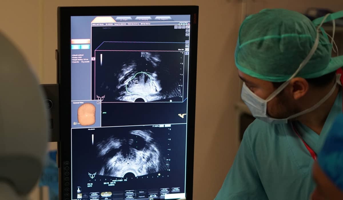
We explain what the grade in the biopsy report means in our article what is the Gleason score, and how an MRI-guided targeted biopsy is performed in our article on MRI-TRUS fusion prostate biopsy.
When this information is combined, the disease is divided into three main risk groups. The intermediate-risk group is itself split into “favourable” and “unfavourable”; the EAU 2026 guideline has formalised this distinction and introduced a new five-tier classification.
| Risk group | Typical definition | General approach |
|---|---|---|
| Low risk | PSA < 10, ISUP 1, T1–T2a | Active surveillance is the standard approach |
| Favourable intermediate risk | ISUP 2 (Gleason 3+4) and PSA < 10 and fewer than half of the biopsy cores positive; limited grade 4 pattern | Active surveillance in selected patients; otherwise surgery or radiotherapy. Focal therapy only within a research setting |
| Unfavourable intermediate risk | ISUP 3 (Gleason 4+3) or PSA 10–20 or T2b–c, or more than one intermediate-risk feature | Surgery (or radiotherapy) |
| High risk | PSA > 20 or ISUP 4–5 or T3 | Surgery; often combined with additional treatments |
Why is life expectancy decisive?
Prostate cancer progresses slowly; the survival benefit of a treatment often becomes apparent only after ten years. For this reason the EAU defines a life expectancy of more than ten years as a prerequisite for benefiting from local treatment. Age alone is not the measure: in patients over 70, overall health is assessed with geriatric assessment tools such as the G8. In patients with a life expectancy below ten years and no symptoms, “watchful waiting” is preferred to avoid the side effects of treatment; unlike active surveillance, it aims not to cure but to control symptoms.
Additional decision tools
When the decision remains uncertain, two supporting tools can come into play. Genomic tests such as Decipher, Prolaris and Oncotype estimate the risk of progression from gene activity in the tumour tissue; according to UCSF data presented at the 2026 AUA congress, a high Decipher score roughly doubles the risk of upgrading to ISUP 3 in patients under surveillance. The EAU considers these tests “promising” but has not yet adopted them for routine use. PSMA PET imaging, on the other hand, is the most sensitive method for showing whether the disease has spread in the body in high-risk patients.
Active Surveillance: When Is “Waiting” the Right Treatment?
Active surveillance does not mean leaving the cancer untreated; it means following the cancer closely with a regular, predefined programme without losing the opportunity for cure. The aim is to avoid the side effects of surgery or radiation, such as urinary incontinence and loss of sexual function, for a cancer that would never cause a problem. The EAU 2026 guideline states this with the strongest level of recommendation: active surveillance should be offered as the standard approach to all low-risk patients and to selected favourable intermediate-risk patients. NCCN 2026 likewise defines active surveillance as the single “strongly recommended” preferred option for low risk. You can also find the basic principles of the method in our article on active surveillance in prostate cancer.
Who are the candidates?
- ISUP 1 (Gleason 3+3) cancer, PSA below 10, tumour confined to the prostate on examination and MRI: for these patients active surveillance is the first option.
- ISUP 2 (Gleason 3+4), but with a limited grade 4 pattern (below 10%), PSA below 10, low PSA density and few positive biopsy cores: surveillance can also be considered in these patients. PRIAS, the world's largest surveillance programme based in the Netherlands, accepts ISUP 2 patients with PSA up to 20 and PSA density up to 0.25 if there is no cribriform or intraductal pattern.
- Cribriform or intraductal pattern, grade ISUP 3 or higher, extension beyond the capsule: if any of these findings is present, active surveillance is not recommended.
How is follow-up done?
The minimum programme recommended by the EAU is: PSA at least every six months, a digital rectal examination once a year, MRI when needed and a repeat biopsy every two to three years, until life expectancy falls below ten years. If the diagnosis was made with an MRI-guided targeted biopsy, the “confirmatory biopsy” in the first year can be omitted. In low-risk patients whose MRI is unchanged and whose PSA density stays below 0.15, the repeat biopsy can also be omitted. This means that surveillance is not equally intensive for every patient and becomes progressively less frequent in low-risk patients.
When is treatment started?
The common principle of the guidelines is that the decision should be based not on a PSA rise or a change on MRI, but on the result of a repeat biopsy. PSA doubling in less than three years or progression on MRI is a reason for a biopsy, not a reason for treatment. Upgrading to ISUP 3, the appearance of a cribriform or intraductal pattern, or a marked increase in tumour volume on biopsy are the strongest reasons to switch to treatment. It is known that patients whose grade rises during surveillance do better than patients diagnosed with the same grade at first diagnosis; in other words, the risk of “missing the opportunity” with surveillance is not as great as feared.
Anxiety is also a medical reason
In about 10% of patients under surveillance, the anxiety of being under constant monitoring reaches a significant level. The EAU recognises this as a valid reason to switch to treatment, but recommends offering psychological support first. Our experience also shows that a patient who has understood the true nature of the disease experiences active surveillance not as waiting but as a conscious choice.
Focal Therapy: Is It Possible to Target Only the Tumour?
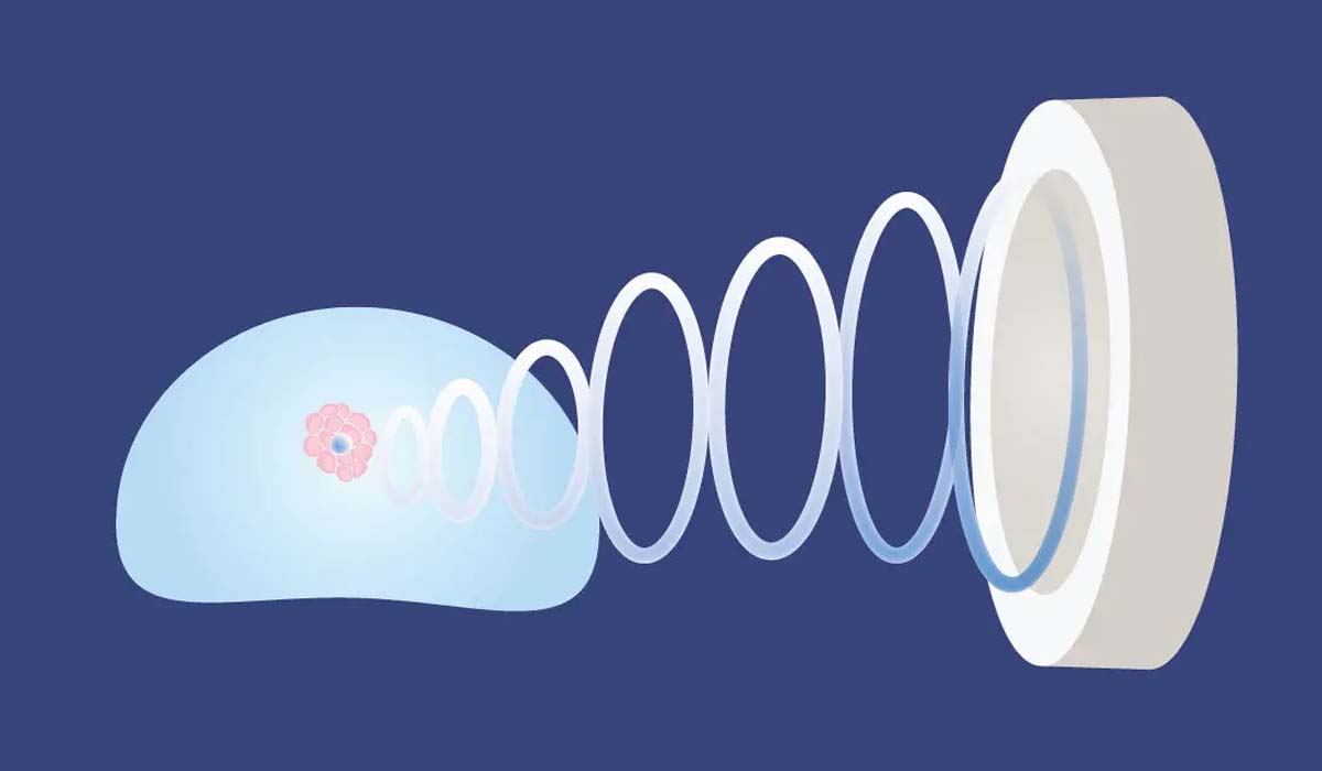
Focal therapy aims to destroy only the area containing the cancer, rather than the whole prostate, using high-intensity focused ultrasound (HIFU), freezing (cryotherapy), an electric field (IRE) or a similar form of energy. The logic is simple: if the location of the tumour can now be seen on MRI, it should be possible to remove only the diseased tissue while preserving the nerves, the urinary sphincter muscle and the urethra. Functional results confirm this expectation: one year after focal HIFU, only a 9% decline in sexual function scores is observed, whereas in treatments that destroy the whole prostate this figure rises to 43%; the continence rate is above 95%.
What do the guidelines say?
Both guidelines approach focal therapy with caution and state this clearly.
- EAU 2026 addresses focal therapy under the heading of “investigational treatments”. It states that the quality of evidence is low, that studies are mostly single-centre and non-comparative, and that there is no standard pathway for patient selection and follow-up. As a result, it recommends that focal therapy be performed within clinical trials or prospective registry programmes.
- NCCN 2026 has added a new section to its guideline this year called “principles of focal therapy”. It emphasises that the standard approach in low-risk patients is active surveillance and asks that focal therapy be approached with care in newly diagnosed patients. In intermediate-risk patients it recommends focal therapy only within a clinical trial, preferably one comparing it with standard treatment. In high-risk patients it stresses that results are likely to be worse than standard treatment, because in these patients the cancer may also be present outside the treated area.
What do the data show?
The largest data set on focal therapy comes from the HEAT and ICE registries in the United Kingdom. The latest analysis, published in July 2026, evaluated 3,477 patients from 14 centres (most treated with HIFU); 48% of the patients were in the favourable intermediate, 23% in the unfavourable intermediate and 25% in the high-risk group. At ten years, death from prostate cancer was 0.13% and metastasis 3.3%; however, over the same period 33% of patients had needed repeat focal treatment and 30% radical treatment such as surgery or radiotherapy. In the authors' own words, up to two sessions of focal therapy can be considered as an option alongside radical treatment in “well-selected” patients; however, the study is observational, meaning the patients were not randomised.
What is missing?
A large randomised trial directly comparing focal therapy with surgery or radiotherapy has not yet been published. The three-year results of the FARP study in Norway have only been presented as a congress abstract. To fill this gap, the 306-patient PART trial in the United Kingdom and the 356-patient ENFORCE trial in the Netherlands are ongoing; both aim to prove that focal therapy is not worse than radical treatment in intermediate-risk patients. Until the results arrive, focal therapy should remain an option applied in patients who meet certain conditions, with its limits understood and with a commitment to regular MRI and biopsy follow-up.
The suitable candidate profile can be summarised as follows: a single, well-defined tumour on MRI, ISUP 2 (selected ISUP 3) grade, PSA below 20, demonstration by both targeted and systematic biopsy that the tumour is not present in the rest of the prostate, and a patient who gives high priority to preserving function. In low-risk patients active surveillance, and in high-risk patients radical treatment, is superior to focal therapy.
Surgery: When the Disease Threatens Life

Surgery, that is, removal of the prostate (radical prostatectomy), is the main treatment aimed at eliminating the cancer completely; radiation therapy (radiotherapy) is the other option used for the same purpose. Guidelines define these two paths as the standard treatment for unfavourable intermediate-risk and high-risk patients with a life expectancy of more than ten years. In the favourable intermediate-risk group, active surveillance, surgery and radiotherapy are presented as options of equal weight; the decision is made together according to the patient's priorities.
Who needs surgery?
- Intermediate-risk patients with grade ISUP 3 or higher, PSA above 10, or a tumour involving a significant part of the prostate.
- All high-risk patients: PSA above 20, grade ISUP 4–5 or extension beyond the capsule.
- Patients in whom progression is detected on biopsy during active surveillance.
- Patients who meet the surveillance criteria but choose treatment after an informed decision.
Surgery: robotic radical prostatectomy
The prostate is removed together with its capsule and the seminal vesicles; the bladder is then reconnected to the urethra. Compared with open surgery, robotic surgery provides less bleeding, a shorter hospital stay and earlier urinary control; in ten-year comparisons with laparoscopic surgery, the “quality” of continence and sexual function was found to be higher with the robotic approach. Nerve-sparing technique, intraoperative frozen-section examination (NeuroSAFE) and preservation of urethral length are methods that aim to preserve function without compromising cancer control. In high-risk patients an extended lymph node dissection is added to the operation. One of the most important advantages of surgery is that the removed tissue can be examined completely and any additional treatment can be planned accordingly. We describe the steps of the operation in our article on robotic surgery for prostate cancer.
Radiotherapy
Image-guided intensity-modulated radiation therapy is the modern standard method. Hypofractionated schedules that shorten the treatment time are recommended by EAU 2026 for intermediate-risk patients; in the PACE-B trial, five-session stereotactic treatment was not inferior to the conventional schedule, with a 95.8% disease-free rate at five years. Hormone therapy is added to radiotherapy for four to six months in intermediate-risk patients and for two to three years in high-risk patients; in the MARCAP analysis covering 12 randomised trials, this addition significantly improved survival. Giving an additional dose to the main tumour visible on MRI (the FLAME trial) reduced the ten-year recurrence rate from 29% to 14%.
Surgery or radiotherapy?
The ProtecT trial showed that the cancer outcomes of these two paths did not differ at 15 years. The difference lies in the side-effect profile: after surgery, urinary incontinence and loss of sexual function appear earlier and more markedly and partly improve over time; after radiotherapy, bowel and urinary complaints and effects related to hormone therapy are more prominent, and loss of sexual function develops more slowly. Surgery generally comes to the fore in young, healthy patients without urinary complaints; radiotherapy in older patients, those with other illnesses or those with a high surgical risk. The final decision should be made in an assessment involving both urology and radiation oncology specialists.
The Studies That Shape These Decisions
The studies below are the real data on which the principles described above are based. Each was carried out in a different period and in a different patient group, so their results should be read together.
| Study | What it compared | Main result | What it means today |
|---|---|---|---|
| ProtecT (UK, 1,643 patients, 15 years; NEJM 2023) | Active monitoring, surgery and radiotherapy in low–intermediate-risk patients detected by PSA testing | Death from prostate cancer at 15 years about 3% in all three arms (2.2–3.1%); no difference. However, metastasis 9.4% in the monitoring arm vs 4.7–5.0% in the treatment arms; 61% of monitored patients switched to treatment within 15 years | Early treatment does not change survival, but close follow-up reduces metastasis. “Monitoring” in this trial was looser than today's active surveillance (no MRI or repeat biopsy); modern surveillance is safer |
| SPCG-4 (Scandinavia, 695 patients, 23 years) | Surgery vs watchful waiting in the pre-PSA era, mostly in patients with palpable tumours | Surgery reduced cancer death and metastasis; the benefit became clear only after 10 years and was greatest under age 65 | In clinically significant, intermediate–high-risk tumours with long life expectancy, surgery saves lives |
| PIVOT (USA, 731 patients, 19 years) | Surgery vs observation in the PSA era, 42% of patients low risk | Overall no significant difference; no benefit at all in low risk, borderline benefit in intermediate risk (16% reduction in risk of death) | Low-risk cancer causes no problems even without treatment; in intermediate risk, treatment may be meaningful |
| GÖTEBORG-1 (Sweden, 25 years; Eur Urol 2025) | Active surveillance in screen-detected low–intermediate-risk patients | Prostate cancer–specific survival at 25 years 94% (99% in very low risk); at 22 years 38% of patients still untreated. Short PSA doubling time, stage T2 and Gleason 7 were markers of surveillance failure | In correctly selected patients, active surveillance is safe with a quarter-century of data |
| Structured surveillance cohorts (PRIAS, Sunnybrook, Johns Hopkins) | Structured active surveillance in low-risk and selected ISUP 2 patients | Ten-year cancer-specific survival 98–100%; more than a third of patients were reclassified over time and switched to treatment | Switching to treatment during surveillance is not a failure but a designed part of the programme |
| Intermediate-risk meta-analyses (2020–2025) | Low- and intermediate-risk surveillance patients | When ISUP 3 is excluded, no difference in metastasis and survival between low-risk and ISUP 2 patients; when ISUP 3 is included, results at 10 years clearly worsen | Surveillance in ISUP 2 should be selective; surveillance is not recommended in ISUP 3 |
| HEAT / ICE registries (UK, 3,477 patients; Eur Urol 2026) | Focal HIFU and cryotherapy | At 10 years cancer death 0.13%, metastasis 3.3%; however 33% repeat focal treatment, 30% radical treatment | Focal therapy preserves function, but one in three patients needs additional treatment and randomised evidence is still lacking |
| PACE-B (874 patients, 5 years) | Five-session stereotactic radiotherapy vs conventional radiotherapy | Disease-free rate at five years 95.8% vs 94.6%; the short schedule was not inferior | In suitable intermediate-risk patients, radiotherapy can be completed in days instead of weeks |
| FLAME (571 patients, 10 years) | Additional radiation dose to the main tumour on MRI | Ten-year biochemical disease-free rate rose from 71% to 86%; side effects did not increase | MRI is now part of radiotherapy planning |
The combined message of these studies is clear: for low-risk prostate cancer, early and aggressive treatment does not prolong life; in contrast, in intermediate–high-risk disease, if life expectancy is sufficient, timely radical treatment markedly reduces metastasis and death from cancer. Focal therapy is a promising bridge between the two, but its level of evidence is not yet complete.
At a Glance: Which Path for Which Patient?
| Situation | Recommended approach | Reason |
|---|---|---|
| ISUP 1, PSA < 10, organ-confined, life expectancy > 10 years | Active surveillance (standard) | Risk of cancer death without treatment below 3% at 15 years; treatment side effects are unnecessary |
| ISUP 2, limited grade 4, PSA < 10, low PSA density, small or invisible tumour on MRI, no cribriform pattern | Active surveillance can be considered; surgery and radiotherapy are equal options | In selected patients, surveillance results do not differ from low risk |
| ISUP 2, single and limited tumour on MRI, high priority on preserving function | Focal therapy only within a trial or registry programme; alternatively surveillance or radical treatment | Function is preserved, but the one-in-three need for retreatment and the lack of randomised evidence must be discussed openly |
| ISUP 3, PSA 10–20 or many positive cores | Surgery: robotic prostatectomy (alternative: radiotherapy + short-term hormone therapy) | Under surveillance, the 10-year risk of metastasis and death increases markedly |
| ISUP 4–5, PSA > 20 or tumour beyond the capsule | Surgery: prostatectomy + lymph node dissection (alternative: radiotherapy + long-term hormone therapy); staging with PSMA PET | Radical treatment reduces metastasis and cancer death; focal therapy is not suitable |
| Any risk group, life expectancy < 10 years, no symptoms | Watchful waiting | Other causes take precedence before the survival benefit of treatment can appear |
Conclusion: Three Questions to Ask Your Doctor
In prostate cancer, the right decision begins with correctly measuring the real danger of the disease. Modern MRI, targeted biopsy, PSMA PET and genomic tests allow us increasingly to distinguish the patient who truly needs treatment from the patient who will never need it. The most important message of the 15–25 years of data we have today is that low-risk prostate cancer does not threaten the life of most men and that these patients can be spared the side effects of treatment. The same data also show beyond doubt that timely radical treatment saves lives in intermediate- and high-risk disease.
The information in this article provides a general framework; no patient's decision can be read from a table alone. When your diagnostic report, your MRI images, your general health and your life priorities are evaluated together, the right path for you is determined. When you go to see your doctor, I recommend asking these three questions:
- “What is my risk group, and which findings determined it?”
- “In my situation, what are the expected cancer outcomes and side effects of surveillance, focal therapy and radical treatment?”
- “How much time do I have for this decision?”
In prostate cancer there is almost always enough time to think, to get a second opinion and to reach a decision together. For the first steps after diagnosis, you may also find our article I have just been diagnosed with cancer: what should I do? helpful.
Clinical Note
This article was written and medically reviewed by Prof. Dr. Murat Binbay, a urologist with more than 25 years of experience and over 1,500 robotic operations. The assessments reflect current international guideline recommendations together with our own clinical practice.
The information here is for general guidance only. Because every patient's history, imaging findings and comorbidities differ, a treatment decision can only be made after an examination. You can reach us through our contact page, or read more about our physician on the about us page.
Sources
- EAU–EANM–ESTRO–ESUR–ISUP–SIOG Prostate Cancer Guidelines 2026 — Treatment (uroweb.org)
- Cornford P et al. EAU Guidelines on Prostate Cancer – 2026 Update, Part I. Eur Urol 2026.
- Spratt DE et al. NCCN Guidelines Insights: Prostate Cancer, Version 5.2026. JNCCN 2026;24(5).
- AUA 2026, International Prostate Forum: The Daily Triage of Grade Group 2 Prostate Cancer (UroToday).
- Light A et al. Oncological Outcomes Following Focal HIFU and Cryotherapy: 3477 Patients from the HEAT and ICE Registries. Eur Urol 2026.
- Bryant RJ et al. The PART randomised trial protocol. BJU Int 2026.
- Te Molder LPW et al. ENFORCE focal randomised controlled trial protocol. BMJ Open 2026.
- Ge S et al. HIFU versus RARP: systematic review and meta-analysis. Ann Surg Oncol 2026.
- Hamdy FC et al. Fifteen-Year Outcomes after Monitoring, Surgery, or Radiotherapy for Prostate Cancer (ProtecT). N Engl J Med 2023;388:1547–1558.
- Bill-Axelson A et al. Radical Prostatectomy or Watchful Waiting in Prostate Cancer – 29-Year Follow-up (SPCG-4). N Engl J Med 2018;379:2319–2329.
- Wilt TJ et al. Follow-up of Prostatectomy versus Observation for Early Prostate Cancer (PIVOT). N Engl J Med 2017;377:132–142.
- Palmstedt E et al. Active surveillance for screen-detected low- and intermediate-risk prostate cancer: extended follow-up up to 25 years in the GÖTEBORG-1 trial. Eur Urol 2025;88:373–380.
- Petrelli F et al. Active surveillance in intermediate-risk prostate cancer: a contemporary synthesis of evidence. Clin Genitourin Cancer 2025;23:102407.
- Shee K et al. A high Decipher genomic risk score is associated with major pathological progression in patients undergoing active surveillance. Eur Urol Oncol 2026.
Last reviewed: 8 October 2026 · Reviewed by: Prof. Dr. Murat Binbay, Urology and Robotic Surgery Specialist
Let Prof. Dr. Murat Binbay's team review your case
If you read this far, you probably have questions. Fill in the two-minute digital consultation form and we will get back to you within 24 hours.
- Answer within 24 hours
- Your data stays confidential

 Önceki Yazı
Testicular Cancer Symptoms: How to Do a Self-Examination and When to See a Urologist
Önceki Yazı
Testicular Cancer Symptoms: How to Do a Self-Examination and When to See a Urologist
 Sonraki Yazı
Which Biopsy Should Be Preferred to Diagnose Prostate Cancer? Why Does Biopsy Quality Matter?
Sonraki Yazı
Which Biopsy Should Be Preferred to Diagnose Prostate Cancer? Why Does Biopsy Quality Matter?
Yorumlar
Yorum Yazın
Henüz yorum yapılmamış. İlk yorumu siz yapın!