Every decision made in the diagnosis of prostate cancer — surveillance, focal therapy or surgery — rests on a single foundation: the biopsy. A biopsy does not only tell us whether cancer is present; it also tells us where it is, how widespread it is and how aggressive it is. If any of this information is missing or wrong, every decision that follows is built on an incomplete or faulty foundation. That is why the question “how was the biopsy done?” is just as important as “was a biopsy done?”. In this article I explain which biopsy approach we prefer today and how biopsy quality determines your treatment options.
Why Is a Biopsy Performed and What Does It Tell Us?
A raised PSA, an examination finding or a suspicious area on MRI suggests that there may be cancer in the prostate; however, cancer can only be diagnosed with a biopsy. During a biopsy, small tissue samples are taken from the prostate with fine needles and examined under the microscope by a pathologist. We explain what the PSA value means in detail in our article what is the PSA test.
A good biopsy report answers three questions:
- Is there cancer?
- How aggressive is it? This is expressed by the Gleason score or the ISUP grade group (from 1 to 5). For details, see our article what is the Gleason score.
- Where is it and how widespread? In samples from which areas cancer was found, in how many samples, and what percentage of each sample contains cancer.
The third question is often overlooked; yet it is precisely this question that determines the treatment options. I will return to it in detail below.
First Principle: MRI First, Then Biopsy
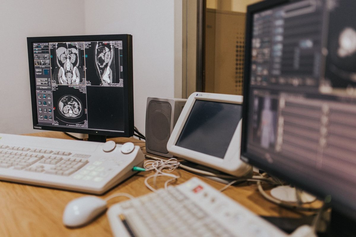
Until recently, prostate biopsy was performed “blind”: under ultrasound guidance, 10–12 random samples were taken from certain areas of the prostate in the hope that the cancer would be hit by these samples. This method could miss significant cancers while unnecessarily detecting insignificant ones.
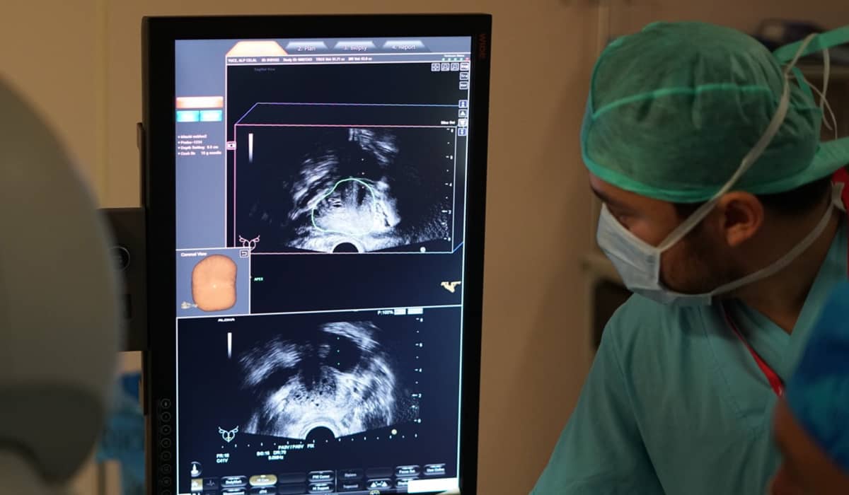
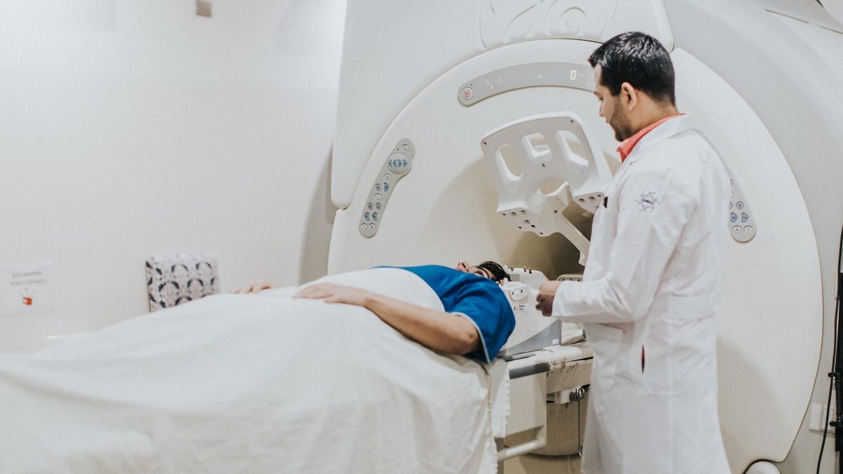
Today the approach has changed. A multiparametric prostate MRI is performed before the biopsy. The MRI shows suspicious areas within the prostate, and these areas are graded with a suspicion score from 1 to 5 (PI-RADS). During the biopsy, the MRI image is superimposed on the live ultrasound image on a computer, and the needle is guided directly into the suspicious area. This is called “MRI-ultrasound fusion biopsy” or “targeted biopsy”. You can find the technical details of the method in our article on MRI-TRUS fusion prostate biopsy.
The most important study demonstrating the value of this approach is the randomised PRECISION trial, carried out in 23 centres in 11 countries and including 500 patients. In this trial, the MRI-first approach detected clinically significant cancer in 38% of patients, compared with 26% for standard blind biopsy. At the same time, the detection of insignificant cancers that do not require treatment fell from 22% to 9%, and 28% of the patients whose MRI was clear avoided biopsy altogether. In other words, the MRI-first pathway both finds more significant cancers and makes fewer unnecessary diagnoses.
In recent years, high-resolution micro-ultrasound technology has also entered this field. In the multicentre randomised OPTIMUM trial published in 2025, micro-ultrasound-guided biopsy was not inferior to MRI fusion biopsy in detecting significant cancers (47% vs 43%). When used together with MRI, this technology is gaining value as a complement that increases the precision of targeting.
Second Principle: Targeted Sampling Alone Is Not Enough — Systematic Mapping Is Essential
MRI is a very valuable guide, but it is not perfect. Some cancers are not visible on MRI, and some lesions are larger than they appear. For this reason, sending a needle only to the target on MRI is not sufficient; the rest of the prostate must also be sampled systematically.
The most important evidence on this subject comes from a prospective study at the US National Cancer Institute that included 2,103 patients, each of whom underwent both targeted and systematic biopsy. Using the two methods together provided an additional cancer diagnosis in about 10% of patients compared with using either alone. Even more importantly, in patients who later had surgery, upgrading of the biopsy grade at surgical pathology — that is, the biopsy showing the cancer as milder than it really was — occurred in 17% with systematic biopsy alone, 9% with targeted biopsy alone, and only 3.5% when both were performed together.
The meaning of this figure is this: an incomplete biopsy hides the true aggressiveness of the cancer in one in every six patients. A well-mapped biopsy reduces this risk to one in thirty.
Third Principle: The Route of the Biopsy — The Transperineal Approach
The biopsy needle can reach the prostate by two routes: through the bowel wall (transrectal) or through the skin area called the perineum, between the anus and the testicles (transperineal).
The transrectal route was used as the standard for many years; however, passing the needle through the bowel can carry bacteria into the prostate and lead to infection. In the transperineal route, the needle enters through sterile skin and the bowel is not touched.
In the randomised PREVENT trial published in 2024, no infections occurred with transperineal biopsy performed without antibiotics, whereas with transrectal biopsy performed with antibiotics, 1.6% of patients developed an infection requiring treatment; cancer detection rates were similar. Another advantage of the transperineal route is easier access to the front parts of the prostate — an area that is difficult to reach via the transrectal route and where some cancers hide.
In our clinic we perform prostate biopsy via the transperineal route, with MRI fusion guidance and under anaesthesia. Our patients are discharged the same day.
Optimal Mapping: The Safeguard of Active Surveillance and Focal Therapy
This section is the most important part of the article.
Today, surgery is not recommended to every patient with prostate cancer. In low-risk patients active surveillance, and in selected intermediate-risk patients focal therapy, which treats only the tumour-bearing area of the prostate, can be applied safely. These two options give the patient the chance to preserve urinary control and sexual function. We discuss in detail which option suits which patient in our article active surveillance, focal therapy and surgery: which path for which patient.
However, these two options share a common prerequisite: knowing precisely what is inside the prostate, where it is and how much of it there is.
- The decision for active surveillance rests on the assurance that “there is only low-grade, low-volume cancer in the prostate; there is no more aggressive focus anywhere else”. This assurance can only be given by a biopsy in which all regions of the prostate have been sampled systematically. In an incompletely sampled prostate, a “low-risk” diagnosis may in fact be “intermediate risk that has not yet been found”.
- The decision for focal therapy is even more delicate: one needs to be able to say “the cancer is only in this area; the rest of the prostate is clean”. The boundaries of the area to be treated, and the fact that the area not to be treated is truly clean, can only be shown by a mapping biopsy in which both targeted and systematic sampling are performed and the location of every sample within the prostate is recorded.
In other words: the better the biopsy is mapped, the less aggressive the treatment we can recommend to the patient. A poorly mapped biopsy pushes the doctor, in the face of uncertainty, to stay on the “safe side” — that is, to treat the whole prostate. A well-mapped biopsy, on the other hand, safely opens the doors to active surveillance and focal therapy.
In our clinic, during a mapping biopsy the location of each sample within the prostate is recorded on a schematic map, and the pathology report is interpreted together with this map. If a patient has had a “blind” or only targeted biopsy at another centre and active surveillance or focal therapy is being considered, we may recommend completing or repeating the biopsy before making a decision. This is not about exposing the patient to a second procedure; it is about protecting them from an unnecessary operation or a missed cancer.
The Criteria of a High-Quality Biopsy
| Criterion | Why is it important? |
|---|---|
| High-quality multiparametric MRI before biopsy | Seeing the target; the MRI must be reported by an experienced radiologist |
| Targeted sampling with MRI-ultrasound fusion | Taking more than one sample from the suspicious area |
| Adding systematic sampling | Catching cancers not visible on MRI; determining the grade correctly |
| Transperineal route | Minimising the risk of infection; access to the front of the prostate |
| Recording the location of each sample | A map for planning active surveillance and focal therapy |
| Experienced uropathologist | Reliability of Gleason grading; a second pathology opinion if needed |
| Patient comfort | Appropriate anaesthesia; allowing the procedure to be carried out calmly and carefully |
How Does the Biopsy Process Work?

- Assessment: PSA, examination and MRI findings are evaluated together, and a decision is made on whether a biopsy is needed. In some patients with a clear MRI and no other risk factors, the biopsy can be postponed.
- Preparation: Blood-thinning medicines are reviewed; with the transperineal route, antibiotic preparation is usually not required.
- Procedure: It is performed under anaesthesia and takes about 20–30 minutes. Targeted and systematic samples are taken and their locations are recorded.
- Afterwards: The patient is discharged the same day. Mild bleeding in the urine and semen may be seen for a few weeks; this is expected.
- Results consultation: The pathology report is ready within 5–7 days. The report, the MRI and the map are evaluated together and discussed with the patient face to face; all options are explained with their benefits and costs.
Frequently Asked Questions
Is a biopsy painful?
When it is performed under appropriate anaesthesia, no pain is felt during the procedure. Afterwards, there may be mild tenderness in the perineal area for a few days.
How many samples are taken?
Targeted and systematic samples together usually number between 16 and 24. More important than the number is that the samples represent all regions of the prostate and that their locations are recorded.
Does a biopsy spread cancer?
No. Current scientific data have not produced any evidence that biopsy spreads cancer.
I had a biopsy at another centre; do I need another one?
Not always. The existing biopsy is evaluated together with the MRI. However, if the biopsy was performed without MRI, with the blind method or without systematic sampling, and active surveillance or focal therapy is being considered, completing the biopsy may be recommended so that the decision is safe.
My MRI is clear; do I need a biopsy?
In a significant proportion of patients with a clear MRI, the biopsy can be postponed. However, PSA level, PSA density, family history and examination findings are evaluated together; the decision is individual.
When can I return to normal life after a biopsy?
Most patients return to daily life the next day. It is recommended to wait a few days before heavy exercise and cycling.
Let Us Review Your Reports
If you have a raised PSA, if a suspicious area has been found on your MRI, or if you would like to know whether a biopsy performed at another centre is adequate, you can send us your MRI images and reports. After the Preliminary Assessment, we will arrange a consultation with you. Tel: +90 530 100 90 85
This article is for general information only and does not replace personal medical advice. Diagnosis and treatment decisions are made after an individual assessment with your doctor.
Clinical Note
This article was written and medically reviewed by Prof. Dr. Murat Binbay, a urologist with more than 25 years of experience and over 1,500 robotic operations. The assessments reflect current international guideline recommendations together with our own clinical practice.
The information here is for general guidance only. Because every patient's history, imaging findings and comorbidities differ, a treatment decision can only be made after an examination. You can reach us through our contact page, or read more about our physician on the about us page.
Sources
- Kasivisvanathan V et al. MRI-targeted or standard biopsy for prostate-cancer diagnosis (PRECISION). N Engl J Med 2018;378:1767–1777.
- Ahdoot M et al. MRI-targeted, systematic, and combined biopsy for prostate cancer diagnosis. N Engl J Med 2020;382:917–928.
- Hu JC et al. Transperineal versus transrectal magnetic resonance imaging–targeted and systematic prostate biopsy to prevent infectious complications (PREVENT). JAMA Oncol 2024.
- Kinnaird A et al. Microultrasonography-guided vs MRI-guided biopsy for prostate cancer diagnosis (OPTIMUM). JAMA 2025.
Last reviewed: 8 October 2026 · Reviewed by: Prof. Dr. Murat Binbay, Urology and Robotic Surgery Specialist
Let Prof. Dr. Murat Binbay's team review your case
If you read this far, you probably have questions. Fill in the two-minute digital consultation form and we will get back to you within 24 hours.
- Answer within 24 hours
- Your data stays confidential

 Önceki Yazı
Active Surveillance, Focal Therapy and Surgery in Prostate Cancer: Which Path for Which Patient?
Önceki Yazı
Active Surveillance, Focal Therapy and Surgery in Prostate Cancer: Which Path for Which Patient?
 Sonraki Yazı
You Have Been Diagnosed With Prostate Cancer: Which Stages Will You Go Through at Our Clinic?
Sonraki Yazı
You Have Been Diagnosed With Prostate Cancer: Which Stages Will You Go Through at Our Clinic?
Yorumlar
Yorum Yazın
Henüz yorum yapılmamış. İlk yorumu siz yapın!