Prostate cancer is one of the most frequently seen types of cancer in men, and early diagnosis is of critical importance for treatment success. While traditional prostate biopsy methods can remain inadequate in some situations, advanced technologies such as MRI-TRUS fusion prostate biopsy increase diagnostic accuracy and allow more effective results to be obtained.
What Is a Prostate Biopsy?
A prostate biopsy is a procedure aiming to detect cancer or other abnormal conditions by taking tissue samples from the prostate gland. It is generally applied in the event of prostate specific antigen (PSA) levels being high or of suspicious findings being detected on rectal examination.
In traditional biopsy methods, random tissue samples are taken from specific areas of the prostate accompanied by transrectal ultrasound (TRUS). However, this method carries the risk of missing the cancer in the event that the tumour is located in small or hard-to-reach areas.
What Is MRI-TRUS Fusion Prostate Biopsy?
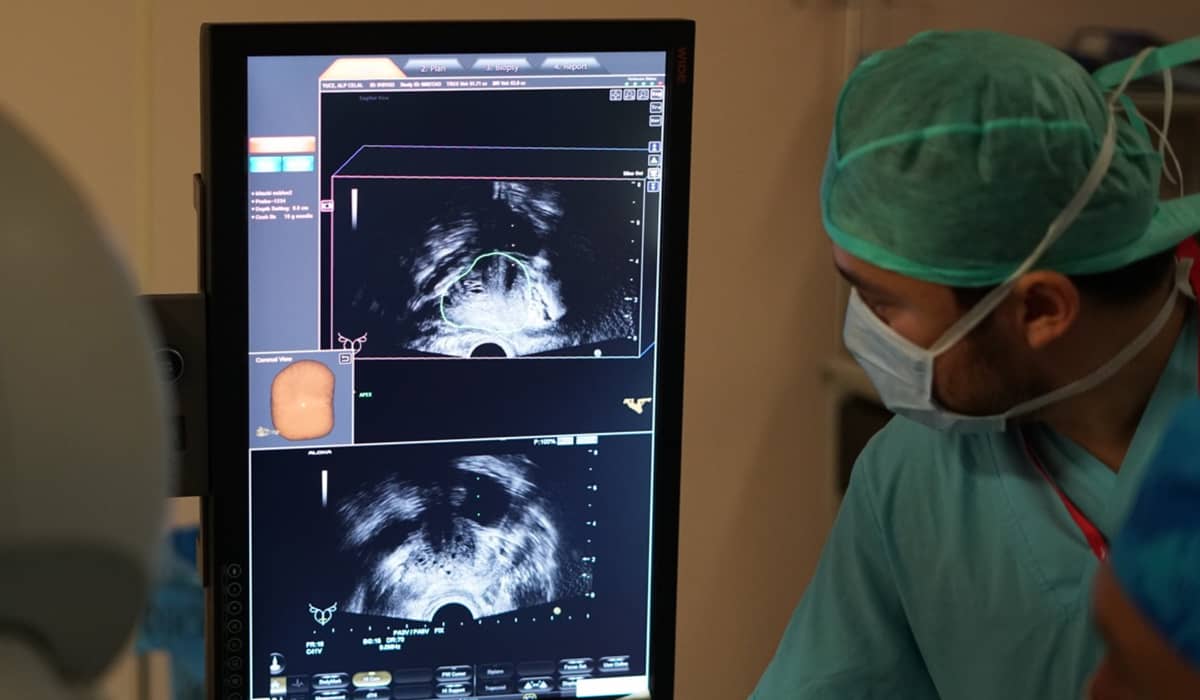
MRI-TRUS fusion prostate biopsy is a highly sensitive biopsy method carried out with the combination of magnetic resonance (MR) imaging and transrectal ultrasonography (TRUS) technologies. Unlike traditional biopsy techniques, this method allows cancerous tissues to be detected more accurately by carrying out targeted sampling.
In this biopsy method, MR images of the prostate are first taken and these images are analysed in detail and the suspicious areas are determined. Because MR scans can show abnormalities in the prostate tissue at high resolution, they play an important role in detecting suspicious areas.
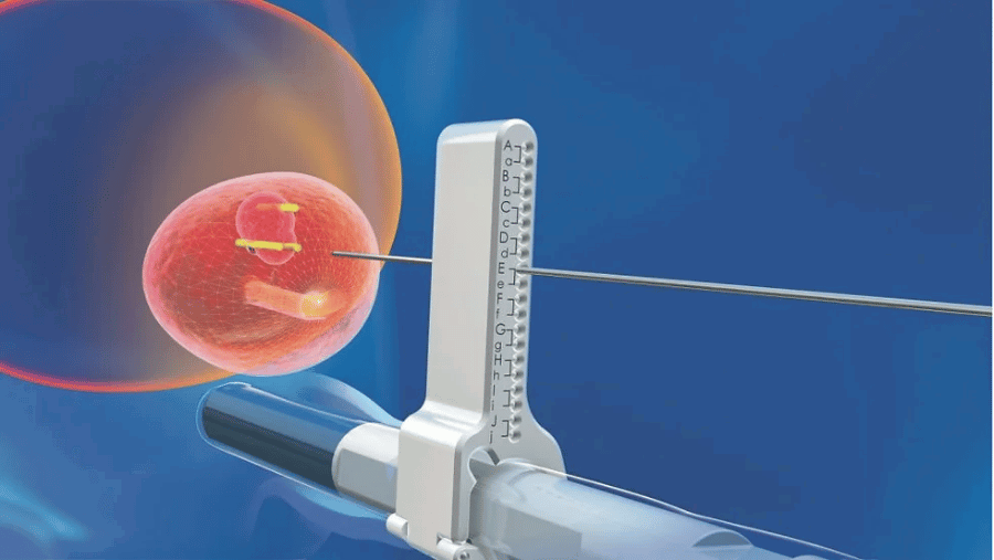
The MR images obtained are then combined with ultrasound images taken in real time with the TRUS device (fusion technology). With the aid of special software these two images are superimposed and a precise guide is created for directing the biopsy needle. In this way, instead of taking random tissue samples during the biopsy procedure, more precise and reliable results are obtained by focusing directly on the suspicious areas.
The Advantages of MRI-TRUS Fusion Biopsy
-
High accuracy rate: this method has a higher cancer detection rate than traditional biopsies. With MRI-TRUS fusion biopsy the diagnostic rate can rise above 95%.
-
Targeted sampling: thanks to the MR images, suspicious areas are clearly identified and the biopsy procedure focuses on these specific areas, thereby reducing unnecessary tissue sampling.
-
Reduced risk of complications: thanks to targeted biopsy, the number of tissue samples taken decreases, and this in turn lowers the risks of complications such as infection.
-
A decrease in the need for repeat biopsy: thanks to the high sensitivity, false negative results and consequently the necessity for repeat biopsy decrease.
How Is the Prostate Biopsy Procedure Carried Out?
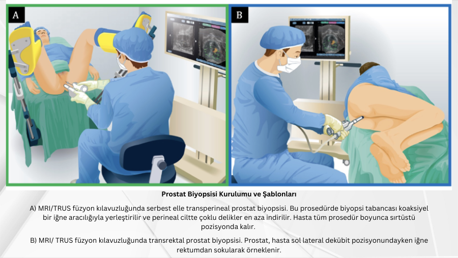
A prostate biopsy is a procedure that allows small samples to be taken from the prostate tissue for the diagnosis of cancer or other abnormal conditions. While random sampling is carried out in traditional biopsy methods, thanks to MRI-TRUS fusion biopsy suspicious areas are precisely identified and targeted tissue can be taken. This method makes the early diagnosis of cancer easier while also reducing the risk of unnecessary biopsy.
The procedure consists of a few basic steps and is planned carefully with patient comfort taken into consideration:
-
MR imaging: firstly, magnetic resonance (MR) images of the prostate are taken. These images reveal abnormalities in the prostate tissue in detail and help in identifying the suspicious areas. MR imaging provides a great advantage particularly in detecting small and hard-to-reach tumours.
-
Combining the images (fusion): the detailed images obtained with MR are combined with the real-time images of the transrectal ultrasonography (TRUS) device used during the biopsy. Thanks to special software these two images are superimposed and it is precisely determined which areas the needle needs to be directed to during the biopsy. This stage prevents unnecessary tissue sampling by allowing a targeted biopsy to be performed.
-
The biopsy procedure: using the fusion images, the biopsy needle is directed straight to the suspicious areas and tissue samples are taken. In this way there is no need for the patient to undergo unnecessarily excessive biopsy and the procedure is carried out more effectively. The tissue samples taken are sent for pathological examination and the presence of cancer or other abnormal cells is investigated.
MRI-TRUS fusion prostate biopsy, by offering more reliable and precise results compared with traditional methods, makes a great contribution to the early diagnosis and treatment process.
MRI-TRUS fusion prostate biopsy offers an important innovation in the early and accurate diagnosis of prostate cancer. With its advantages such as a high accuracy rate, targeted sampling and a reduced risk of complications, this method stands out as a more effective option compared with traditional biopsy methods. For patients with suspected prostate cancer, the use of this advanced technology allows more precise and reliable data to be obtained in treatment planning.
Clinical Note
This article was written and medically reviewed by Prof. Dr. Murat Binbay, a urologist with more than 25 years of experience and over 1,500 robotic operations. The assessments reflect current international guideline recommendations together with our own clinical practice.
The information here is for general guidance only. Because every patient's history, imaging findings and comorbidities differ, a treatment decision can only be made after an examination. You can reach us through our contact page, or read more about our physician on the about us page.
Sources
- European Association of Urology — Prostate Cancer Guidelines
- National Cancer Institute (NIH) — Prostate Cancer
- Türk Üroloji Derneği — Güncel Kılavuzlar
Last reviewed: 8 September 2026 · Reviewed by: Prof. Dr. Murat Binbay, Urology and Robotic Surgery Specialist
Let Prof. Dr. Murat Binbay's team review your case
If you read this far, you probably have questions. Fill in the two-minute digital consultation form and we will get back to you within 24 hours.
- Answer within 24 hours
- Your data stays confidential

Urology and Robotic Surgery Specialist · 25+ years of experience
Last medically reviewed:
 Önceki Yazı
What Is the Gleason Score?
Önceki Yazı
What Is the Gleason Score?
 Sonraki Yazı
Why Does the PSA Level Rise After Prostate Cancer Treatment?
Sonraki Yazı
Why Does the PSA Level Rise After Prostate Cancer Treatment?
Yorumlar
Yorum Yazın
Henüz yorum yapılmamış. İlk yorumu siz yapın!