Retroperitoneal fibrosis is a rare disease characterized by abnormal inflammation and increased connective tissue in the retroperitoneal space located at the back of the abdominal cavity. The resulting fibrotic tissue often develops around major blood vessels and the ureters. Entrapment or external compression of the ureters within this tissue restricts the flow of urine produced by the kidneys to the bladder, potentially leading to hydronephrosis and loss of kidney function.
While the disease progresses slowly and silently in some individuals, it may present in others with lower back or flank pain, impaired kidney function, or recurrent urinary tract issues. The primary goal of treatment in retroperitoneal fibrosis is not only to control the existing fibrotic tissue, but also to protect the kidneys from irreversible damage caused by ureteral obstruction.
A significant proportion of retroperitoneal fibrosis cases are idiopathic, meaning no definitive cause can be demonstrated. However, it is now known that in certain cases, the disease may be linked to the immune system and specifically to a systemic fibroinflammatory condition called IgG4-related disease. In addition, certain medications, malignancies, radiotherapy, past surgical procedures, and various inflammatory conditions can cause secondary retroperitoneal fibrosis.
What is the Retroperitoneum and Where Does Retroperitoneal Fibrosis Occur?
The retroperitoneum is the anatomical region situated behind the peritoneal lining of the abdomen. The kidneys, a major portion of the ureters, the aorta, the inferior vena cava, as well as numerous vessels, nerves, and lymphatic structures reside in this area.
In retroperitoneal fibrosis, an inflammatory and fibrotic tissue layer typically develops around major blood vessels, particularly the aorta. Over time, this tissue can extend toward surrounding structures. Because the ureters pass through this region, they are among the structures most significantly impacted by the disease.
Unilateral or bilateral entrapment of the ureters within the fibrotic tissue can prevent urine from passing from the kidney to the bladder. Consequently, the condition is evaluated not only as a rheumatologic or inflammatory issue, but also as a critical urologic disease.
What Causes Retroperitoneal Fibrosis?
The precise cause of retroperitoneal fibrosis cannot be identified in every patient. Therefore, the disease is generally classified into two main categories: idiopathic retroperitoneal fibrosis and secondary retroperitoneal fibrosis.
Idiopathic Retroperitoneal Fibrosis
Cases where no specific underlying cause can be detected are termed idiopathic. Today, chronic inflammatory processes triggered by the immune system are believed to play a central role in a significant portion of these patients.
Over time, persistent inflammation leads to dense connective tissue formation and fibrosis, resulting in the encasement of retroperitoneal structures such as the ureters and major vessels.
IgG4-Related Retroperitoneal Fibrosis
In some patients, retroperitoneal fibrosis may manifest as part of IgG4-related disease. IgG4-related disease is a systemic condition capable of causing inflammation and fibrosis across various organ systems.
Affected individuals may exhibit involvement in the pancreas, salivary glands, lacrimal glands, kidneys, major vessels, and other organ systems. Alternatively, retroperitoneal fibrosis can present without involvement of other organs.
Elevated serum IgG4 levels can support the diagnosis but are not individually diagnostic. A definitive evaluation relies on a combination of clinical findings, imaging results, and tissue biopsy when necessary.
Secondary Retroperitoneal Fibrosis
Less frequently, retroperitoneal fibrosis develops secondary to an underlying condition or external factor. Secondary causes include:
- Long-term use of specific medications
- Prior radiation therapy to the abdominal or pelvic region
- Previous major abdominal surgeries
- Certain chronic infections
- Lymphoma and other malignancies
- Inflammatory conditions located in the retroperitoneal space
- Specific systemic autoimmune diseases
Distinguishing secondary forms is critical because treatment must target the underlying etiology in addition to managing the fibrosis itself.
Who is Affected by Retroperitoneal Fibrosis?
Retroperitoneal fibrosis is a rare condition that predominantly presents in middle-aged to older adults. It occurs more frequently in men than in women, though it can develop across different age groups and in female patients as well.
No single risk factor accounts for the onset of the disease. Immune system disorders, IgG4-related disease, specific pharmaceuticals, prior radiation or surgical history, and underlying malignancies are all carefully considered during patient evaluation.
What Are the Symptoms of Retroperitoneal Fibrosis?
The symptoms of retroperitoneal fibrosis generally correlate with the progression of the disease and the degree of extrinsic compression on the ureters. Early-stage manifestations can be quite subtle and non-specific.
Primary symptoms may include:
- Lower back pain
- Flank or side pain
- Abdominal pain
- Dull pain radiating to the groin
- Fatigue and malaise
- Loss of appetite
- Unexplained weight loss
- Low-grade fever
- Nausea
- Decreased urine output
- Impaired renal function
- Recurrent urinary tract infections
Because ureteral obstruction often develops gradually, some patients may not experience acute flank pain. In such cases, retroperitoneal fibrosis is discovered incidentally when hydronephrosis is detected during ultrasonography or computed tomography performed for unrelated reasons.
How Does Retroperitoneal Fibrosis Affect the Ureters?
Urine produced by the kidneys is transported to the bladder through two narrow muscular tubes called ureters. In retroperitoneal fibrosis, these ureters can become encased or externally compressed by the surrounding dense fibrotic tissue.
This entrapment impedes the flow of urine from the kidney to the bladder. As backpressure builds above the site of narrowing, the kidney's collecting system dilates—a condition known as hydronephrosis.
If the obstruction persists over an extended period, renal parenchyma can sustain progressive damage, compromising the kidney's filtration capacity. When both ureters are affected, renal function deterioration is typically more pronounced.
Can Retroperitoneal Fibrosis Cause Kidney Failure?
Yes. Severe ureteral compression and long-standing urinary tract obstruction can lead to a decline in renal function. Particularly when the disease affects both ureters, elevated serum creatinine levels along with acute or chronic renal failure may develop.
Consequently, establishing the extent of renal impairment is a central priority during diagnosis. In patients with significantly compromised kidney function or marked ureteral obstruction, medical therapy alone may be insufficient; prompt decompression of the urinary drainage system is often required first.
How is Retroperitoneal Fibrosis Diagnosed?
Retroperitoneal fibrosis cannot be diagnosed via a single laboratory test. Diagnosis requires a synthesis of clinical symptoms, renal function parameters, inflammatory markers, and advanced imaging findings.
Blood and Urine Tests
Blood tests are conducted to evaluate renal performance through serum creatinine and related parameters. Inflammatory markers such as Erythrocyte Sedimentation Rate (ESR) and C-Reactive Protein (CRP) provide objective data regarding disease activity.
Depending on clinical features, serum IgG4 measurements, autoimmune panels, and additional laboratory investigations may be indicated. However, none of these laboratory values independently confirms or excludes the diagnosis.
Ultrasonography
Ultrasonography is primarily valuable for identifying the presence and severity of hydronephrosis. However, detailed visualization of the fibrotic mass boundaries and its anatomical relationship to major abdominal vessels requires cross-sectional imaging modalities.
Computed Tomography (CT)
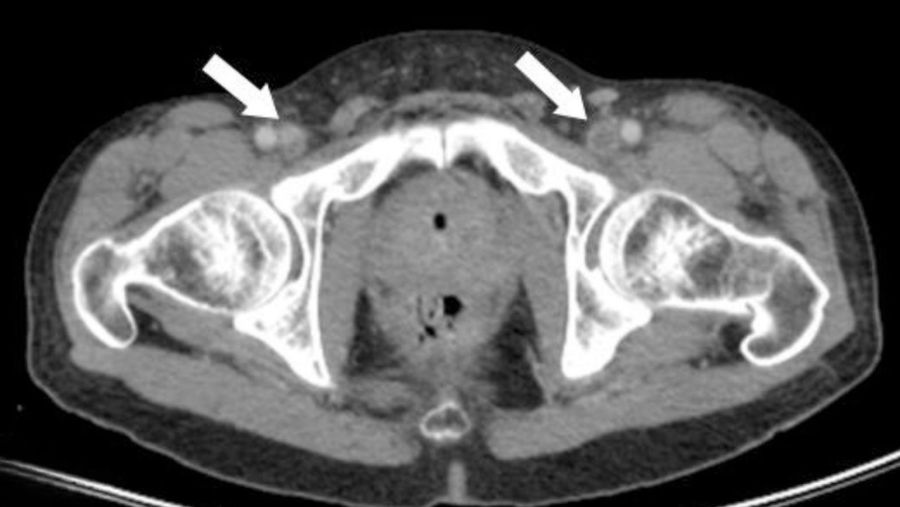
Contrast-enhanced CT is one of the most widely utilized modalities for assessing the distribution of retroperitoneal fibrosis and its relation to adjacent structures. It allows precise evaluation of ureteral involvement, renal pelvicalyceal dilation, and retroperitoneal tissue extent.
Magnetic Resonance Imaging (MRI)
MRI is valuable for delineating the anatomical extent of fibrotic tissue and monitoring treatment response. It serves as a suitable alternative or complementary study to contrast-enhanced CT in selected patients.
Is a Biopsy Necessary in Retroperitoneal Fibrosis?
Routine tissue biopsy is not mandatory for every patient with retroperitoneal fibrosis. When clinical presentation and imaging findings are highly characteristic, treatment may be initiated directly.
However, a biopsy becomes essential under the following circumstances:
- Atypical imaging features
- Suspicion of underlying malignancy
- Unusual anatomical location of the retroperitoneal mass
- Inadequate response to initial medical therapy
- Need to definitively rule out lymphoma or other neoplasms
- Scenarios where tissue diagnosis significantly alters management strategy
The decision to perform a biopsy is individualized based on radiological patterns and overall clinical context.
What Conditions Can Mimic Retroperitoneal Fibrosis?
Not all soft-tissue masses in the retroperitoneum represent retroperitoneal fibrosis. Lymphoma, metastatic carcinomas, primary retroperitoneal tumors, chronic infections, and various inflammatory conditions can display similar radiological appearances.
Excluding malignant disease is therefore critical, particularly in patients presenting with atypical imaging characteristics. This diagnostic challenge represents one of the primary reasons for pursuing tissue biopsy in uncertain cases.
How is Retroperitoneal Fibrosis Treated?
Treatment of retroperitoneal fibrosis rests on two core objectives: suppressing the active inflammatory and fibrotic disease process, and maintaining renal drainage to safeguard kidney function whenever ureteral obstruction occurs.
Management cannot be limited to a single medication or surgical procedure. Renal function, degree of hydronephrosis, extent of ureteral entrapment, underlying etiology, and immune-mediated factors must be evaluated collaboratively.
Double J Stent Placement for Ureteral Obstruction
When retroperitoneal fibrosis compresses a ureter and impairs renal drainage, a Double J ureteral stent can be inserted. The stent spans from the renal pelvis to the bladder, creating an internal conduit that permits urine to bypass the narrowed segment.
Stenting is particularly useful for restoring immediate urine flow in patients exhibiting declining kidney function. It also serves as a stabilizing measure, providing time for medical therapies to take effect.
However, a stent does not treat the underlying retroperitoneal fibrosis itself; its role is strictly mechanical to ensure ureteral patency. Once the systemic disease is controlled, serial imaging and functional assessments determine whether the stent can be safely removed.
When is Percutaneous Nephrostomy Indicated?
In cases where ureteral stenting is technically unfeasible, obstruction is severe, or immediate decompression is required, a percutaneous nephrostomy may be performed.
Under image guidance, a catheter is inserted directly through the skin into the renal collecting system. This allows urine to drain into an external collection bag, immediately relieving high pressure within the kidney.
Rapid renal decompression via nephrostomy is critical in scenarios involving profound renal insufficiency, anuria, severe electrolyte imbalances, or superinfected obstruction.
Medical Therapy for Retroperitoneal Fibrosis
In idiopathic and immune-mediated retroperitoneal fibrosis, medical therapy aims to suppress active inflammation, halt fibrotic progression, and induce regression of the retroperitoneal mass whenever possible.
Corticosteroid Therapy
Corticosteroids constitute the primary first-line medical intervention for active idiopathic retroperitoneal fibrosis. Dosage and duration are tailored according to disease activity, baseline renal function, radiological features, and patient comorbidities.
During treatment, potential corticosteroid-related side effects—such as hyperglycemia, hypertension, bone density loss, and infection risk—must be closely monitored. Consequently, long-term steroid regimens require structured physician oversight.
Immunosuppressive and Immunomodulatory Agents
In patients who show inadequate response to steroids, suffer disease relapses, or require steroid-sparing strategies, alternative immunosuppressive or immunomodulatory agents are considered.
Medications such as azathioprine, mycophenolate mofetil, and methotrexate may be utilized. In refractory, recurrent, or IgG4-related cases, targeted biologic agents such as rituximab can be integrated into the treatment plan following specialist evaluation.
Because no universal drug regimen fits every scenario, multidisciplinary management involving rheumatology, nephrology, and urology is strongly recommended.
Surgical Treatment Options
Surgical intervention is considered when ureteral obstruction persists despite medical therapy, long-term stenting or nephrostomy becomes unmanageable, or recurrent obstructions occur.
The primary goal of surgery is to meticulously dissect the ureter free from surrounding fibrotic tissue and reposition it to prevent recurrent entrapment. This procedure is known as ureterolysis.
What is Ureterolysis Surgery?
Ureterolysis is the surgical mobilization of the ureter from dense retroperitoneal fibrotic tissue. When necessary, the ureter is transposed or wrapped with protective tissue (such as omentum) to minimize future contact with the fibrotic process.
Key indications for ureterolysis include:
- Persistent ureteral obstruction despite appropriate medical therapy
- Recurrent or unresolving hydronephrosis
- Inability to tolerate long-term ureteral stenting
- Complications associated with indwelling stents or nephrostomies
- Desire for a definitive anatomical solution to obstruction
- Need for open tissue biopsy during surgical management
While ureterolysis addresses local mechanical obstruction, it does not eradicate the systemic inflammatory process driving the disease. Therefore, medical treatment and long-term surveillance are often still required postoperatively.
Robotic Surgery in Retroperitoneal Fibrozis
In suitable candidates, ureterolysis can be performed using laparoscopic or robotic-assisted surgical methods rather than open surgery. Robotic systems provide high-definition 3D visualization and wristed instrumentation, offering technical precision during delicate ureteral dissection out of dense fibrotic tissue.
During robotic ureterolysis, the ureter is freed from surrounding fibrosis and may be transposed or protected using reconstructive techniques adapted to patient anatomy.
Robotic approach is not necessary or suitable for every patient. Disease extent, prior abdominal operations, vascular involvement, and surgical expertise in reconstructive urology guide procedural selection.
What Happens If Retroperitoneal Fibrosis is Left Untreated?
Untreated active retroperitoneal fibrosis can cause progressive ureteral compression, leading to worsening hydronephrosis and permanent loss of renal function.
Furthermore, involvement of retroperitoneal vascular or adjacent structures can cause secondary complications. Management should never be guided by pain symptoms alone, as significant silent structural damage can occur.
Bilateral obstruction, rapidly rising serum creatinine, acute oliguria/anuria, or signs of urinary infection require urgent medical evaluation and intervention.
Does Retroperitoneal Fibrosis Recur?
Retroperitoneal fibrosis can run a chronic relapsing course. Even after achieving clinical remission with medical therapy, disease reactivation remains possible.
Recurrence risk varies based on underlying etiology, association with IgG4-related disease, initial disease extent, treatment responsiveness, and therapeutic modalities employed.
Symptom resolution does not negate the need for ongoing surveillance. Renal function tests, acute-phase reactants, and serial imaging studies should be conducted at routine intervals.
Post-Treatment Follow-Up Protocol
Follow-up strategies are tailored to disease severity and interventions performed. The goal is early detection of disease relapse or recurrent ureteral obstruction.
Standard monitoring includes:
- Serum creatinine and kidney function panels
- Inflammatory markers (CRP and ESR)
- Ultrasonographic monitoring of hydronephrosis
- Periodic CT or MRI cross-sectional imaging
- Stent surveillance in patients with active internal drains
- Evaluation for extra-retroperitoneal organ involvement in IgG4-related disease
Imaging interpretation focuses not only on mass size changes but also on functional renal drainage and ureteral patency.
Is Retroperitoneal Fibrosis Completely Curable?
Disease trajectory varies substantially among individuals. In many patients, inflammation can be controlled effectively with medical therapy, alleviating extrinsic ureteral compression. In others, persistent fibrotic tissue may necessitate long-term stenting, reconstructive surgery, or continuous surveillance.
Prompt diagnosis and early restoration of urinary drainage before irreversible parenchymal damage occurs represent key prognostic factors. Chronic, long-standing renal structural damage may not be fully reversible.
Frequently Asked Questions About Retroperitoneal Fibrosis
Is retroperitoneal fibrosis a form of cancer?
Idiopathic retroperitoneal fibrosis is not cancer. However, certain malignancies can mimic retroperitoneal fibrosis or trigger secondary fibrotic reactions. Excluding underlying neoplasm is an essential step during evaluation.
Is retroperitoneal fibrosis dangerous?
It can cause serious health complications by obstructing ureters and threatening renal function. Prognosis depends on the degree of renal impairment and involvement of adjacent retroperitoneal structures rather than mass size alone.
Does retroperitoneal fibrosis cause ureteral strictures?
Yes. The disease can encase or compress the ureters externally, impeding urine passage and producing functional ureteral strictures, hydronephrosis, and loss of kidney function.
Can retroperitoneal fibrosis affect both kidneys simultaneously?
Yes. Depending on fibrotic extent, one or both ureters can be affected. Bilateral ureteral involvement presents a higher risk of rapid renal function deterioration and demands close management.
Which specialist treats retroperitoneal fibrosis?
Urology plays a central role due to ureteral and renal involvement. However, because it is frequently a systemic inflammatory disorder, care often involves a multidisciplinary team including rheumatology, nephrology, radiology, pathology, and occasionally oncology.
Is a ureteral stent permanent in retroperitoneal fibrosis?
Not necessarily. Stents are frequently used as temporary measures to secure renal drainage. If inflammation recedes following medical therapy and ureteral compression resolves, the stent can often be removed. However, long-term stenting may be required in persistent cases.
Do steroids cure retroperitoneal fibrosis?
Corticosteroids are a cornerstone therapy for suppressing active inflammation in idiopathic or immune-mediated cases. However, patient response must be monitored closely, and medical therapy alone may not suffice if mechanical ureteral obstruction is severe.
Does surgery completely cure retroperitoneal fibrosis?
Ureterolysis resolves local mechanical compression on the ureters, but it does not halt the underlying systemic inflammatory process. Ongoing medical therapy and follow-up are typically required after surgery.
Can retroperitoneal fibrosis relapse?
Yes. Disease reactivation can occur even after successful initial therapy. Regular monitoring of kidney function and serial imaging are necessary to detect relapse early.
When is emergency evaluation required for retroperitoneal fibrosis?
Immediate medical attention is warranted if patients experience a sudden drop in urine volume, anuria, high fever with chills, severe flank pain, rapid deterioration in general health, or acute worsening of kidney function tests. Superinfected urinary obstruction represents a medical emergency requiring urgent renal drainage.
Let Prof. Dr. Murat Binbay's team review your case
If you read this far, you probably have questions. Fill in the two-minute digital consultation form and we will get back to you within 24 hours.
- Answer within 24 hours
- Your data stays confidential

Urology and Robotic Surgery Specialist · 25+ years of experience
Last medically reviewed:


Yorumlar
Yorum Yazın
Henüz yorum yapılmamış. İlk yorumu siz yapın!